. 2021 Jul 27;14(8):734.
doi: 10.3390/ph14080734.

Antibodies Enhance the Suppressive Activity of Extracellular Vesicles in Mouse Delayed-Type Hypersensitivity

Affiliations

Antibodies Enhance the Suppressive Activity of Extracellular Vesicles in Mouse Delayed-Type Hypersensitivity

Katarzyna Nazimek et al. Pharmaceuticals (Basel). .

Abstract

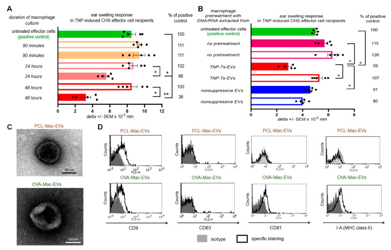
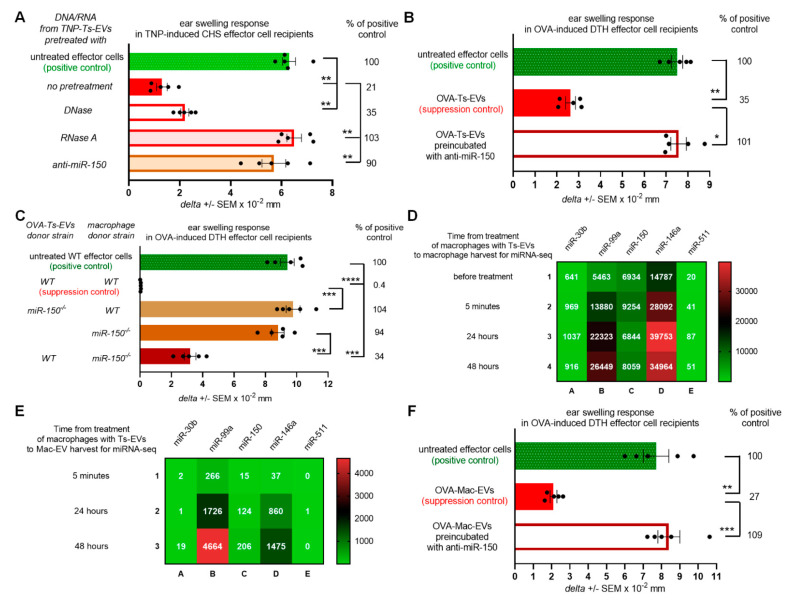
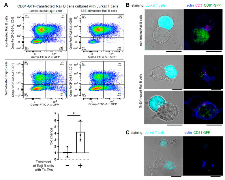
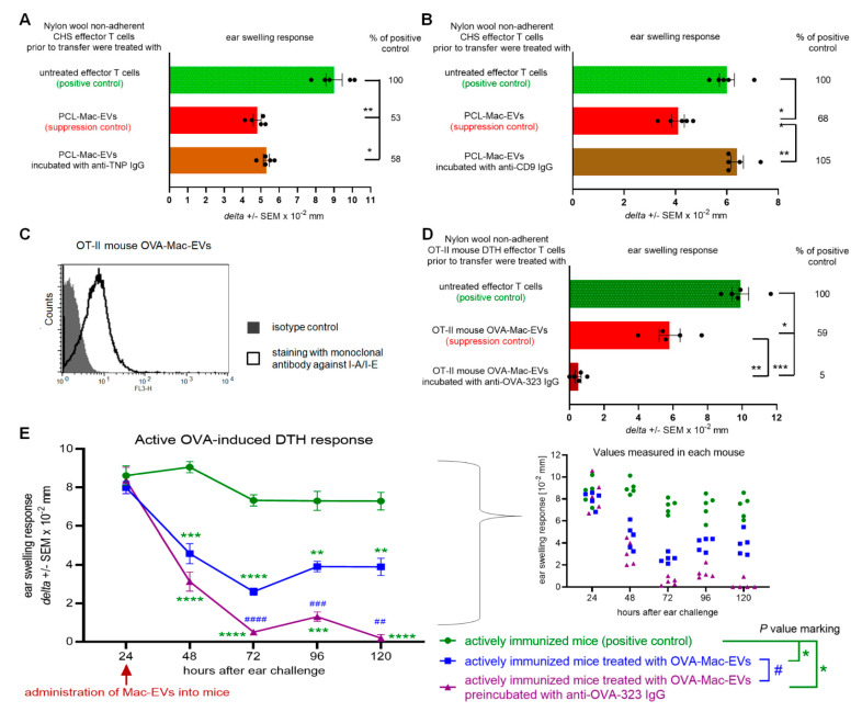
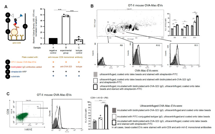
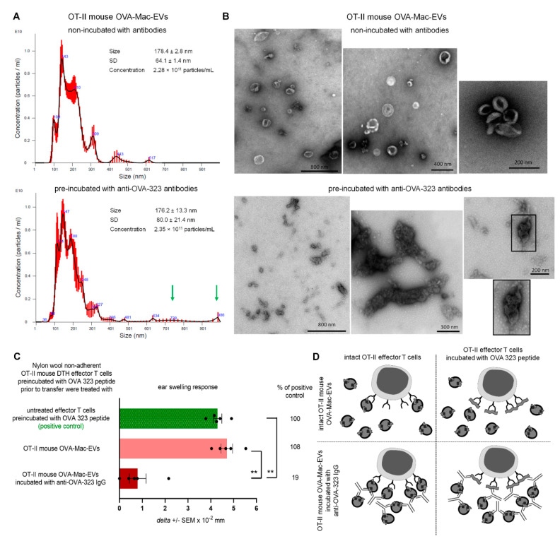
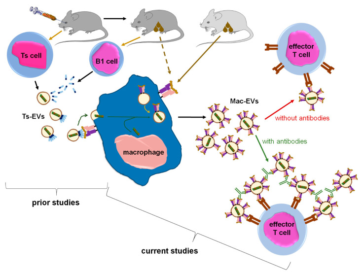
Previously, we showed that mouse delayed-type hypersensitivity (DTH) can be antigen-specifically downregulated by suppressor T cell-derived miRNA-150 carried by extracellular vesicles (EVs) that target antigen-presenting macrophages. However, the exact mechanism of the suppressive action of miRNA-150-targeted macrophages on effector T cells remained unclear, and our current studies aimed to investigate it. By employing the DTH mouse model, we showed that effector T cells were inhibited by macrophage-released EVs in a miRNA-150-dependent manner. This effect was enhanced by the pre-incubation of EVs with antigen-specific antibodies. Their specific binding to MHC class II-expressing EVs was proved in flow cytometry and ELISA-based experiments. Furthermore, by the use of nanoparticle tracking analysis and transmission electron microscopy, we found that the incubation of macrophage-released EVs with antigen-specific antibodies resulted in EVs' aggregation, which significantly enhanced their suppressive activity in vivo. Nowadays, it is increasingly evident that EVs play an exceptional role in intercellular communication and selective cargo transfer, and thus are considered promising candidates for therapeutic usage. However, EVs appear to be less effective than their parental cells. In this context, our current studies provide evidence that antigen-specific antibodies can be easily used for increasing EVs' biological activity, which has great therapeutic potential.

Keywords: antigen-presenting cells; antigen-specific T cell suppression; contact hypersensitivity; delayed-type hypersensitivity; extracellular vesicles; immune tolerance; intercellular communication; macrophages; miRNA-150; therapeutic activity of exosomes.


Conflict of interest statement

K.N. and K.B. declare that they are inventors in a patent application number P.435582 submitted by Jagiellonian University, Krakow, Poland for the method of generating the antibody-aggregated macrophage EVs. The other authors declare no conflict of interest. The funders had no role in the design of the study; in the collection, analyses, or interpretation of data; in the writing of the manuscript, or in the decision to publish the results.

Figures

Figure 1
Figure 1
Macrophages treated with Ts-EVs release inhibitory Mac-EVs that differ in marker’s expression pattern depending on mouse immunizing antigen. (A) Macrophages, after 30 min treatment at 37 °C with TNP-Ts-EVs, were cultured in protein-free MDM medium. Yielded supernatant was collected 90 min, 24 h or 48 h later, filtered and ultracentrifuged, and resulting fractions (i.e., pellet–filled bars; and supernatant above–open bars) were used to treat CHS effector cells prior to their transfer into naive recipients (n = 5 per group) that were immediately challenged with hapten to elicit CHS reaction, measured as ear swelling 24 h later. (B) Untreated macrophages or macrophages treated for 30 min at 37 °C with DNA/RNA extracted from either TNP-Ts-EVs or control, non-suppressive EVs, were cultured in protein-free MDM medium for 48 h. Yielded supernatant was filtered and ultracentrifuged, and resulting fractions (i.e., pellet–filled bars; and supernatant above–open bars) were used to treat CHS effector cells prior to their transfer into naive recipients (n = 5 per group) that were immediately challenged with hapten to elicit CHS reaction, measured as ear swelling 24 h later. (C) PCL-Mac-EVs (produced by TNP-Ts-EV-treated macrophages from PCL-sensitized mice) and OVA-Mac-EVs (produced by OVA-Ts-EV-treated macrophages from OVA-immunized mice) were absorbed onto cupper grid, negatively stained with 3% uranyl acetate, and visualized with TEM microscope. (D) PCL-Mac-EVs and OVA-Mac-EVs were coated onto latex beads, stained with fluoresceinated antibodies against selected EVs’ markers, including CD9, CD63, CD81 tetraspanins and I-A molecules, and analyzed with flow cytometry. Data are expressed as delta ± SEM. One-way ANOVA with post hoc RIR Tukey test; * p < 0.05, ** p < 0.01.
Figure 2
Figure 2
Ts-EV-treated macrophages release Mac-EVs inhibiting DTH in miRNA-150-dependent manner. (A) CHS effector T cells and macrophages were incubated with TNP-Ts-EV-extracted DNA/RNA pretreated with DNase, RNase A or anti-miR-150, and then adoptively transferred to naive recipients (n = 5 per group) that were immediately challenged with hapten to elicit CHS reaction, measured as ear swelling 24 h later. (B) DTH effector T cells and macrophages were incubated with OVA-Ts-EVs, where indicated pretreated with anti-miR-150, and then adoptively transferred to naive recipients (n = 5 per group) that 24 h later were challenged with OVA to elicit DTH reaction, measured as ear swelling 24 h later. (C) DTH effector cells were treated with either OVA-Mac-EVs from wild type mice, EVs from macrophages treated with OVA-Ts-EVs from miRNA-150−/− mice, EVs from untreated miRNA-150−/− mouse macrophages, or with EVs from miRNA-150−/− mouse macrophages treated with OVA-Ts-EVs from wild type mice. Twenty four hours later recipients of adoptively transferred DTH effector cells (n = 5 per group) were challenged with OVA to elicit DTH reaction, measured as ear swelling 24 h later. (D) RNA extracts from untreated macrophages or macrophages treated with OVA-Ts-EVs were subjected to miRNA deep sequencing (n = 1 per time-point). (E) RNA extracts from OVA-Mac-EVs collected from 5 min-, 24 h- or 48 h- macrophage culture supernatant were subjected to miRNA deep sequencing (n = 1 per time-point). (F) DTH effector cells were incubated with OVA-Mac-EVs, where indicated pretreated with anti-miR-150, and then adoptively transferred to naive recipients (n = 5 per group) that 24 h later were challenged with OVA to elicit DTH reaction, measured as ear swelling 24 h later. Data are expressed as delta ± SEM. One-way ANOVA with post hoc RIR Tukey test; * p < 0.05, ** p < 0.01, *** p < 0.005, **** p < 0.001.
Figure 3
Figure 3
OVA-Ts-EVs modulate vesicle-mediated interaction of Raji B cells and Jurkat T cells at the immune synapse. (A) CD81-GFP-transfected Raji B cells were pulsed with SEE superantigen for 30 min at 37 °C and then cultured with Jurkat T cells in the presence of OVA-Ts-EVs. Twenty four hours later, cells were stained with viability dye and fluoresceinated antibodies against CD19, and analyzed with flow cytometry (n = 3). Relative changes in the percentage of CD19negGFPpos Jurkat T cells caused by SEE-stimulation of Raji B cells were calculated as follows: (percentage of CD19negGFPpos events in SEE-stimulated sample)/(percentage of CD19negGFPpos events in unstimulated sample); and shown in the graph. (B) CD81-GFP-transfected Raji B cells were left untreated (upper panel) or were treated with OVA-Ts-EVs for 4 h at 37 °C (lower panel), both populations were then pulsed with SEE for 30 min at 37 °C, and mixed with CMAC-stained Jurkat T cells (1 × 10⁵ cells) in a ratio 1:1. Then, cell mixtures were plated onto Poly-L-Lys-coated slides for 1 h incubation at 37 °C, fixed, blocked, stained with selected primary and then secondary antibodies, mounted on Prolong Gold and analyzed with confocal microscope; scale bar: 10 µm. (C) CD81-GFP-transfected Raji B cells were treated with OVA-Ts-EVs for 4 h at 37 °C, pulsed with SEE for 30 min at 37 °C, and cultured with CMAC-stained Jurkat T cells (1 × 10⁵ cells) in a ratio 1:1 for 24 h on standard culture plate. Then, cell mixtures were plated onto Poly-L-Lys-coated slides for 1 h incubation at 37 °C, fixed, blocked, stained with selected primary and then secondary antibodies, mounted on Prolong Gold and analyzed with confocal microscope; scale bar: 10 µm. Data are expressed as mean ± SD. Two-tailed Student t-test; * p < 0.05.
Figure 4
Figure 4
Antibodies modulate the suppressive activity of PCL- and OVA-Mac-EVs. (A) CHS effector T cells were incubated for 30 min at 37 °C with PCL-Mac-EVs, where indicated pre-incubated with anti-TNP IgG antibodies. Afterwards, CHS effector T cells were adoptively transferred to naive recipients (n = 5 per group) that were immediately challenged with hapten to elicit CHS reaction, measured as ear swelling 24 h later. (B) CHS effector T cells were incubated for 30 min at 37 °C with PCL-Mac-EVs, where indicated pre-incubated with anti-CD9 IgG antibodies. Afterwards, CHS effector T cells were adoptively transferred to naive recipients (n = 5 per group) that were immediately challenged with hapten to elicit CHS reaction, measured as ear swelling 24 h later. (C) OT-II mouse OVA-Mac-EVs were coated onto latex beads, stained with fluoresceinated antibodies against I-A/I-E molecules, and analyzed with flow cytometry. (D) DTH effector T cells from OVA-immunized OT-II mice were incubated for 30 min at 37 °C with OT-II mouse OVA-Mac-EVs, where indicated pre-incubated with anti-OVA-323 IgG antibodies, and then were adoptively transferred to naive wild type recipients (n = 5 per group) that 24 h later were challenged with OVA to elicit DTH reaction, measured as ear swelling 24 h later. (E) After measuring of 24-h DTH ear swelling, actively immunized C57BL/6 mice (n = 5 per group) were administered intraperitoneally with OT-II mouse OVA-Mac-EVs alone or preincubated with anti-OVA-323 IgG antibodies, and subsequent DTH ear swelling was measured up to 120 h after challenge. Data are expressed as delta ± SEM. One-way or two-way ANOVA with post hoc RIR Tukey test; * p < 0.05, ** p < 0.01, *** p < 0.005, **** p < 0.001, # p < 0.05, ## p < 0.01, ### p < 0.005, #### p < 0.001.
Figure 5
Figure 5
Anti-OVA-323 antibodies specifically bind to OVA-Mac-EVs from OT-II mice. (A) OT-II mouse OVA-Mac-EVs were incubated on ELISA plate coated with anti-CD9 antibodies for 2 h at room temperature. Then, biotinylated anti-OVA-323 IgG or biotinylated isotype IgG were added to selected wells, and the plate was incubated overnight at 4 °C. Afterwards, streptavidin-HRP was added to each well, which was followed by 45 min incubation at room temperature and addition of TMB substrate. Reaction was stopped after 5 min with 1 M H3PO4, and absorbance was measured at 450 nm with a reference wavelength of 570 nm. Then, blanked absorbance values were normalized to mean blanked absorbance detected in wells with added OVA-Mac-EVs but not antibodies (n = 4). (B) OT-II mouse OVA-Mac-EVs were either incubated with biotinylated anti-OVA-323 IgG overnight at 4 °C, ultracentrifuged, and coated onto latex beads, or coated onto latex beads and incubated with biotinylated anti-OVA-323 IgG for an hour at room temperature. Then, both OVA-Mac-EV preparations were incubated with streptavidin-FITC, and analyzed with flow cytometry (n = 3). (C) OT-II mouse OVA-Mac-EVs were incubated with either biotinylated anti-OVA-323 IgG, or FITC-conjugated isotype IgG overnight at 4 °C. After ultracentrifugation, both OVA-Mac-EV preparations were coated onto latex beads, incubated with streptavidin-FITC and fluoresceinated antibodies against CD9 and I-A/I-E, and analyzed with flow cytometry (n = 3). Data are expressed as mean ± SD. One-way or two-way ANOVA with post hoc RIR Tukey test; * p < 0.05, ** p < 0.01, *** p < 0.005.
Figure 6
Figure 6
Incubation of OT-II mouse OVA-Mac-EVs with anti-OVA-323 antibodies leads to their aggregation and enhances their suppressive activity. (A) OT-II mouse OVA-Mac-EVs were incubated alone or with biotinylated anti-OVA-323 IgG overnight at 4 °C. After ultracentrifugation, both OVA-Mac-EV preparations were subjected to nanoparticle tracking analysis (NTA). (B) OT-II mouse OVA-Mac-EVs were incubated alone or with biotinylated anti-OVA-323 IgG overnight at 4 °C. After ultracentrifugation, both OVA-Mac-EV preparations were absorbed onto cupper grid, negatively stained with 3% uranyl acetate, and visualized with TEM microscope. (C) OT-II mouse OVA-Mac-EVs were incubated alone or with biotinylated anti-OVA-323 IgG overnight at 4 °C. After ultracentrifugation, both OVA-Mac-EV preparations were used for 30 min treatment at 37 °C of OT-II mouse DTH effector T cells that had been pre-incubated with OVA-323 peptide for 20 min at 37 °C. Then, DTH effector T cells were adoptively transferred to naive wild type recipients (n = 5 per group) that 24 h later were challenged with OVA to elicit DTH reaction, measured as ear swelling 24 h later. (D) Scheme showing the proposed mechanism of enhancement of OVA-Mac-EVs’ suppressive activity against OVA-specific DTH effector T cells, in some instances pre-incubated with OVA-323 peptide, induced by anti-OVA-323 IgG antibodies. NTA results are shown as mean ± SD, and DTH ear swellings are expressed as delta ± SEM. One-way ANOVA with post hoc RIR Tukey test; ** p < 0.01.
Figure 7
Figure 7
Summary of the findings in the context of existing knowledge. From prior studies we knew that intravenous tolerization of mice with antigen-coupled syngeneic red blood cells induces Ts cells to release miRNA-150 in Ts-EVs that are then coated with antigen-specific antibody light chains. The latter are secreted by B1 cells activated by antigen applied to mice via cutaneous route. Subsequently, Ts-EVs target macrophages that are able to present antigenic determinant complexed with MHC class II after cutaneous immunization of donor animal. In turn, Ts-EV-targeted macrophages suppress CHS and DTH responses in mice. Currently, we have shown that macrophages treated with Ts-EV-transmitted miRNA-150 begin to release Mac-EVs that also contain miRNA-150, which finally inhibits CHS and DTH effector T cell activity. Crucially, Mac-EVs express MHC class II molecules that are likely complexing the antigenic determinant, which enables the specific targeting of effector T cells, and also binding of specific IgG antibodies by Mac-EVs. The latter finding allowed us to discover that antigen-specific antibodies aggregate Mac-EVs, which greatly enhances their suppressive activity against effector T cells. Thus, one can conclude that macrophages multiply the number of miRNA-150 copies and release them in MHC class II-positive EVs to amplify the suppressive effect and direct it against specific effector T cells, and that this effect is significantly enhanced by incubating Mac-EVs with antigen-specific antibodies.

Similar articles

Cited by

References

    1. Maas S.L.N., Breakefield X.O., Weaver A.M. Extracellular vesicles: Unique intercellular delivery vehicles. Trends Cell Biol. 2017;27:172–188. doi: 10.1016/j.tcb.2016.11.003. - DOI - PMC - PubMed
    1. Sedgwick A.E., D’Souza-Schorey C. The biology of extracellular microvesicles. Traffic. 2018;19:319–327. doi: 10.1111/tra.12558. - DOI - PMC - PubMed
    1. Kalluri R., LeBleu V.S. The biology, function, and biomedical applications of exosomes. Science. 2020;367:eaau6977. doi: 10.1126/science.aau6977. - DOI - PMC - PubMed
    1. Gaurav I., Thakur A., Iyaswamy A., Wang X., Chen X., Yang Z. Factors affecting extracellular vesicles based drug delivery systems. Molecules. 2021;26:1544. doi: 10.3390/molecules26061544. - DOI - PMC - PubMed
    1. Bryniarski K., Ptak W., Jayakumar A., Püllmann K., Caplan M.J., Chairoungdua A., Lu J., Adams B.D., Sikora E., Nazimek K., et al. Antigen-specific, antibody-coated, exosome-like nanovesicles deliver suppressor T-cell microRNA-150 to effector T cells to inhibit contact sensitivity. J. Allergy Clin. Immunol. 2013;132:170–181.e9. doi: 10.1016/j.jaci.2013.04.048. - DOI - PMC - PubMed