

Canine Myocytes Represent a Good Model for Human Ventricular Cells Regarding Their Electrophysiological Properties
- PMID: 34451845
- PMCID: PMC8398821
- DOI: 10.3390/ph14080748
Canine Myocytes Represent a Good Model for Human Ventricular Cells Regarding Their Electrophysiological Properties
Abstract
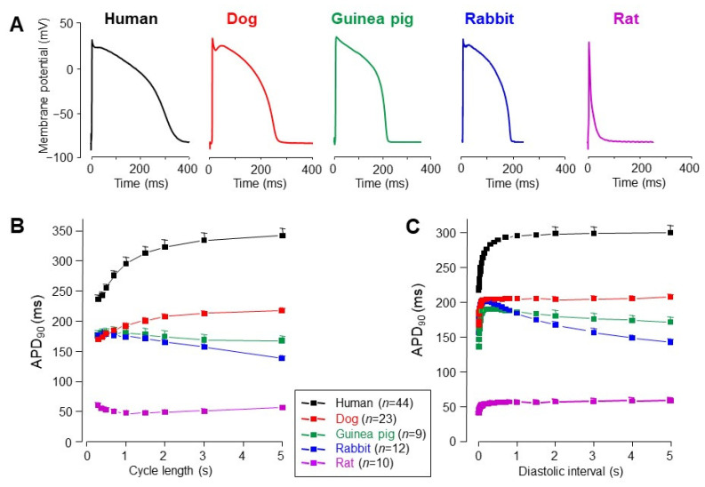
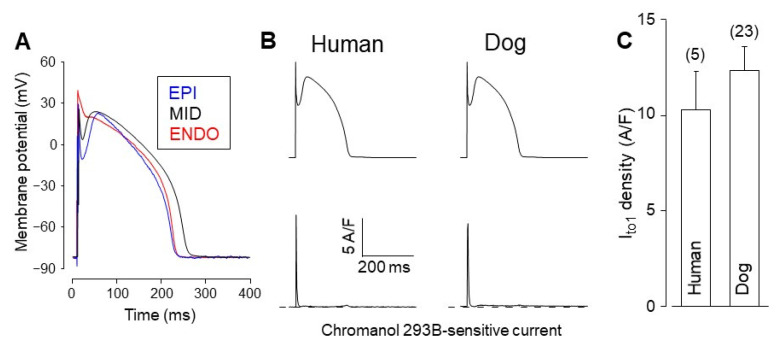
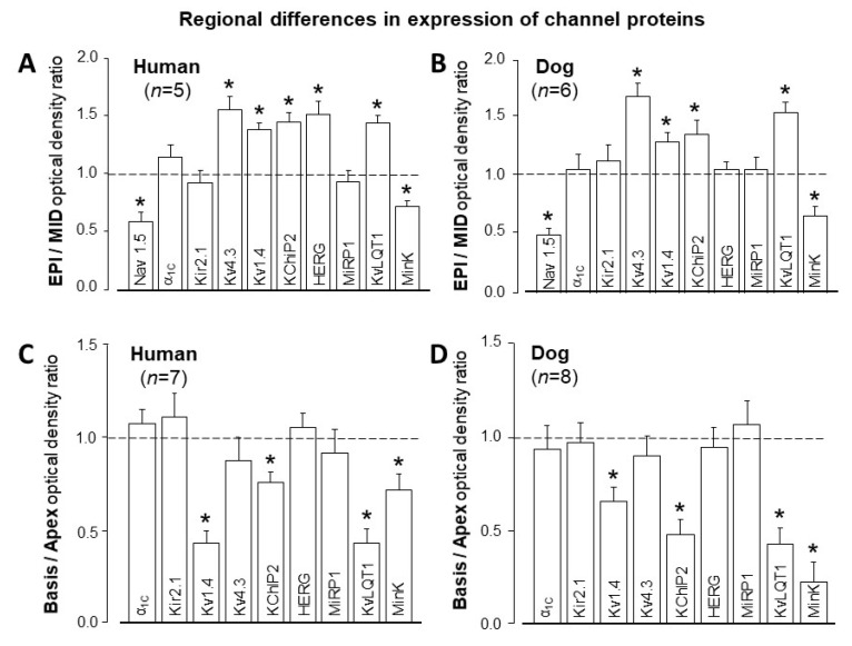
Due to the limited availability of healthy human ventricular tissues, the most suitable animal model has to be applied for electrophysiological and pharmacological studies. This can be best identified by studying the properties of ion currents shaping the action potential in the frequently used laboratory animals, such as dogs, rabbits, guinea pigs, or rats, and comparing them to those of human cardiomyocytes. The authors of this article with the experience of three decades of electrophysiological studies, performed in mammalian and human ventricular tissues and isolated cardiomyocytes, summarize their results obtained regarding the major canine and human cardiac ion currents. Accordingly, L-type Ca2+ current (ICa), late Na+ current (INa-late), rapid and slow components of the delayed rectifier K+ current (IKr and IKs, respectively), inward rectifier K+ current (IK1), transient outward K+ current (Ito1), and Na+/Ca2+ exchange current (INCX) were characterized and compared. Importantly, many of these measurements were performed using the action potential voltage clamp technique allowing for visualization of the actual current profiles flowing during the ventricular action potential. Densities and shapes of these ion currents, as well as the action potential configuration, were similar in human and canine ventricular cells, except for the density of IK1 and the recovery kinetics of Ito. IK1 displayed a largely four-fold larger density in canine than human myocytes, and Ito recovery from inactivation displayed a somewhat different time course in the two species. On the basis of these results, it is concluded that canine ventricular cells represent a reasonably good model for human myocytes for electrophysiological studies, however, it must be borne in mind that due to their stronger IK1, the repolarization reserve is more pronounced in canine cells, and moderate differences in the frequency-dependent repolarization patterns can also be anticipated.
Keywords: action potential configuration; action potential voltage clamp; canine myocytes; cardiac ion currents; human ventricular cells.
Conflict of interest statement
The authors declare no conflict of interest.
Figures


Similar articles
-
Ion current profiles in canine ventricular myocytes obtained by the "onion peeling" technique.J Mol Cell Cardiol. 2021 Sep;158:153-162. doi: 10.1016/j.yjmcc.2021.05.011. Epub 2021 Jun 3. J Mol Cell Cardiol. 2021. PMID: 34089737
-
Ionic mechanisms limiting cardiac repolarization reserve in humans compared to dogs.J Physiol. 2013 Sep 1;591(17):4189-206. doi: 10.1113/jphysiol.2013.261198. Epub 2013 Jul 22. J Physiol. 2013. PMID: 23878377 Free PMC article.
-
Effects of pioglitazone on cardiac ion currents and action potential morphology in canine ventricular myocytes.Eur J Pharmacol. 2013 Jun 15;710(1-3):10-9. doi: 10.1016/j.ejphar.2013.03.047. Epub 2013 Apr 12. Eur J Pharmacol. 2013. PMID: 23588116
-
The impact of single cell voltage clamp on the understanding of the cardiac ventricular action potential.Cardioscience. 1992 Sep;3(3):131-44. Cardioscience. 1992. PMID: 1384746 Review.
-
Cardiac voltage-gated ion channels in safety pharmacology: Review of the landscape leading to the CiPA initiative.J Pharmacol Toxicol Methods. 2017 Sep;87:11-23. doi: 10.1016/j.vascn.2017.04.002. Epub 2017 Apr 11. J Pharmacol Toxicol Methods. 2017. PMID: 28408211 Review.
Cited by
-
ABT-333 (Dasabuvir) Increases Action Potential Duration and Provokes Early Afterdepolarizations in Canine Left Ventricular Cells via Inhibition of IKr.Pharmaceuticals (Basel). 2023 Mar 25;16(4):488. doi: 10.3390/ph16040488. Pharmaceuticals (Basel). 2023. PMID: 37111245 Free PMC article.
-
Comparative analysis of the ten Tusscher and Tomek human ventricular cell models at cellular, tissue, and organ levels: Implications for post-infarct ventricular tachycardia simulation.Physiol Rep. 2025 Jul;13(13):e70435. doi: 10.14814/phy2.70435. Physiol Rep. 2025. PMID: 40641123 Free PMC article.
-
Cardiac electrophysiological remodeling associated with enhanced arrhythmia susceptibility in a canine model of elite exercise.Elife. 2023 Feb 23;12:e80710. doi: 10.7554/eLife.80710. Elife. 2023. PMID: 36815557 Free PMC article.
-
Spatiotemporal patterns of early afterdepolarizations underlying abnormal T-wave morphologies in a tissue model of the Purkinje-ventricular system.PLoS One. 2023 Jan 9;18(1):e0280267. doi: 10.1371/journal.pone.0280267. eCollection 2023. PLoS One. 2023. PMID: 36622850 Free PMC article.
-
Guidelines for assessment of cardiac electrophysiology and arrhythmias in small animals.Am J Physiol Heart Circ Physiol. 2022 Dec 1;323(6):H1137-H1166. doi: 10.1152/ajpheart.00439.2022. Epub 2022 Oct 21. Am J Physiol Heart Circ Physiol. 2022. PMID: 36269644 Free PMC article. Review.
References
-
- Pueyo E., Dangerfield C.E., Britton O.J., Virág L., Kistamás K., Szentandrássy N., Jost N., Varró A., Nánási P.P., Burrage K., et al. Experimentally-based computational investigation into beat-to-beat variability in ventricular repolarization and its response to ionic current inhibition. PLoS ONE. 2016;11:e0151461. doi: 10.1371/journal.pone.0151461. - DOI - PMC - PubMed
-
- Tomek J., Bueno-Orovio A., Passini E., Zhou X., Minchole A., Britton O., Bartolucci C., Severi S., Shrier A., Virag L., et al. Development, calibration, and validation of a novel human ventricular myocyte model in health, disease, and drug block. eLife. 2019;8:e48890. doi: 10.7554/eLife.48890. - DOI - PMC - PubMed
Publication types
Grants and funding
LinkOut - more resources
Full Text Sources
Miscellaneous
