. 2021 Aug 12;14(8):792.
doi: 10.3390/ph14080792.

Chirality-Dependent Anti-Inflammatory Effect of Glutathione after Spinal Cord Injury in an Animal Model

Affiliations

Chirality-Dependent Anti-Inflammatory Effect of Glutathione after Spinal Cord Injury in an Animal Model

Seong-Jun Kim et al. Pharmaceuticals (Basel). .

Abstract

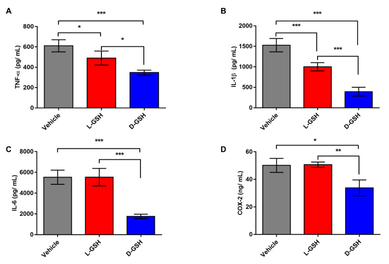
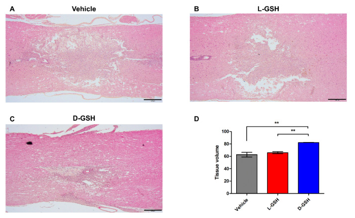
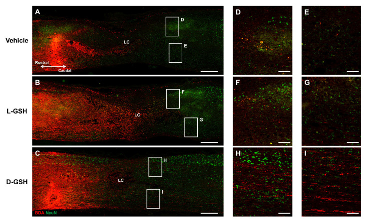
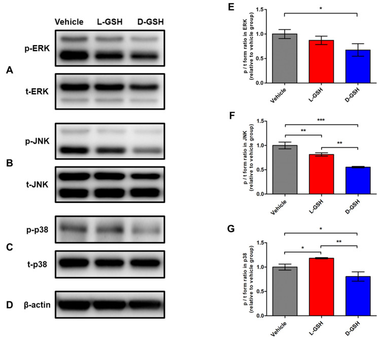
Neuroinflammation forms a glial scar following a spinal cord injury (SCI). The injured axon cannot regenerate across the scar, suggesting permanent paraplegia. Molecular chirality can show an entirely different bio-function by means of chiral-specific interaction. In this study, we report that d-chiral glutathione (D-GSH) suppresses the inflammatory response after SCI and leads to axon regeneration of the injured spinal cord to a greater extent than l-chiral glutathione (L-GSH). After SCI, axon regrowth in D-GSH-treated rats was significantly increased compared with that in L-GSH-treated rats (*** p < 0.001). Secondary damage and motor function were significantly improved in D-GSH-treated rats compared with those outcomes in L-GSH-treated rats (** p < 0.01). Moreover, D-GSH significantly decreased pro-inflammatory cytokines and glial fibrillary acidic protein (GFAP) via inhibition of the mitogen-activated protein kinase (MAPK) signaling pathway compared with L-GSH (*** p < 0.001). In primary cultured macrophages, we found that D-GSH undergoes more intracellular interaction with activated macrophages than L-GSH (*** p < 0.001). These findings reveal a potential new regenerative function of chiral GSH in SCI and suggest that chiral GSH has therapeutic potential as a treatment of other diseases.

Keywords: chirality; glial scars; glutathione; neuroinflammation; spinal cord injuries.


Conflict of interest statement

The authors declare no conflict of interest.

Figures

Figure 1
Figure 1
Circular dichroism (CD) spectra of L-chiral form of GSH (L-GSH; red) and D-chiral form of GSH (D-GSH; blue).
Figure 2
Figure 2
The protein secretion levels in the injured spinal cord segments of the vehicle, L-GSH, and D-GSH groups after SCI. Quantification of (A) Tumor necrosis factor-α (TNF-α), (B) interleukin (IL)-1β, (C) IL-6, and (D) cyclooxygenase-2 (COX-2). Results are the mean ± SEM; * p < 0.05, ** p < 0.01, and *** p < 0.001; one-way ANOVA with Tukey post hoc test.
Figure 3
Figure 3
Immunofluorescence analysis in composite tiled scans of sagittal sections stained for glial scar (anti-glia fibrillary acidic protein, GFAP, Green), inflammatory cytokine (anti-tumor necrosis factor-α, TNF-α, Red). Proximal border (PB) and distal border (DB) away from the lesion center (LC). (A) Representative merged image for GFAP and TNF-α in the vehicle group. (B) Representative image for GFAP in the L-GSH group. (C) Higher magnification of the proximal border (boxed area) from (B). (D) Representative image for GFAP in the D-GSH group. (E) Higher magnification of the proximal border (boxed area) from (D). (F) Representative image for TNF-α in the L-GSH group. (G) Higher magnification of the proximal border (boxed area) from (F). (H) Representative image for TNF-α in the D-GSH group. (I) Higher magnification of the proximal border (boxed area) from (H). Scale bar, 500 μm (A,B,D,F,H) and 100 μm (C,E,G,I). Quantitative analyses of the fluorescence intensity for (J) GFAP and (K) TNF-α. Results are the mean ± standard error of the mean (SEM); * p < 0.05, ** p < 0.01, and *** p < 0.001; one-way ANOVA with Tukey post hoc test.
Figure 4
Figure 4
Histopathological changes were evaluated for the (A) vehicle-, (B) L-GSH-, and (C) D-GSH-treated rats. Representative images of the spinal cord stained with H&E in the vehicle, L-GSH, and D-GSH groups. (D) Quantitative analysis of the tissue volume. Results are the mean ± standard error of the mean (SEM), ** p < 0.01; one-way ANOVA with Tukey post hoc test.
Figure 5
Figure 5
BDA-labelled axons in composite tiled scans of sagittal sections stained for neuron (anti-NeuN, Green), axon (anti-streptavidin, Red). LC, lesion center. Representative images of the (A) vehicle group, (B) L-GSH group, and (C) D-GSH group after SCI in rats. (D,E) Higher magnification of boxed areas past the lesion center from (A). (F,G) Higher magnification of boxed areas past the lesion center from (B). (H,I) Higher magnification of boxed areas past the lesion center from (C). Scale bar, 500 μm (AC) and 100 μm (DI).
Figure 6
Figure 6
The phosphorylation activities of the MAPK signaling pathway in the vehicle, L-GSH, and D-GSH groups. Representative images of the p and t forms of (A) ERK, (B) JNK, (C) p38, and (D) β-actin. Quantitative analyses of the p/t forms of (E) ERK, (F) JNK, and (G) p38. The p/t form volume in the vehicle group was set to 1-fold, and the ratio was relatively calculated and quantified. Results are the mean ± SEM; * p < 0.05, ** p < 0.01, and *** p < 0.001; one-way ANOVA with Tukey post hoc test.

Similar articles

Cited by

References

    1. Jain N.B., Ayers G.D., Peterson E.N., Harris M.B., Morse L., O’Connor K.C., Garshick E. Traumatic spinal cord injury in the United States, 1993–2012. JAMA. 2015;313:2236–2243. doi: 10.1001/jama.2015.6250. - DOI - PMC - PubMed
    1. Rust R., Kaiser J. Insights into the Dual Role of Inflammation after Spinal Cord Injury. J. Neurosci. 2017;37:4658–4660. doi: 10.1523/JNEUROSCI.0498-17.2017. - DOI - PMC - PubMed
    1. Ko W.K., Kim S.J., Jo M.J., Choi H., Lee D., Kwon I.K., Lee S.H., Han I.B., Sohn S. Ursodeoxycholic Acid Inhibits Inflammatory Responses and Promotes Functional Recovery After Spinal Cord Injury in Rats. Mol. Neurobiol. 2019;56:267–277. doi: 10.1007/s12035-018-0994-z. - DOI - PubMed
    1. Kim J.W., Mahapatra C., Hong J.Y., Kim M.S., Leong K.W., Kim H.W., Hyun J.K. Functional Recovery of Contused Spinal Cord in Rat with the Injection of Optimal-Dosed Cerium Oxide Nanoparticles. Adv. Sci. 2017;4:1700034. doi: 10.1002/advs.201700034. - DOI - PMC - PubMed
    1. Qian T., Guo X., Levi A.D., Vanni S., Shebert R.T., Sipski M.L. High-dose methylprednisolone may cause myopathy in acute spinal cord injury patients. Spinal Cord. 2005;43:199–203. doi: 10.1038/sj.sc.3101681. - DOI - PubMed

LinkOut - more resources