Review
. 2021 Aug 23;14(8):829.
doi: 10.3390/ph14080829.

Extracellular Vesicles: A Double-Edged Sword in Sepsis

Affiliations
Review

Extracellular Vesicles: A Double-Edged Sword in Sepsis

Marlies Burgelman et al. Pharmaceuticals (Basel). .

Abstract

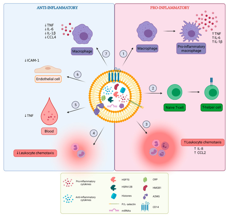
Sepsis is defined as a life-threatening organ dysfunction caused by a dysregulated host response to an infection. Several studies on mouse and patient sepsis samples have revealed that the level of extracellular vesicles (EVs) in the blood is altered compared to healthy controls, but the different functions of EVs during sepsis pathology are not yet completely understood. Sepsis EVs are described as modulators of inflammation, lymphocyte apoptosis, coagulation and organ dysfunction. Furthermore, EVs can influence clinical outcome and it is suggested that EVs can predict survival. Both detrimental and beneficial roles for EVs have been described in sepsis, depending on the EV cellular source and the disease phase during which the EVs are studied. In this review, we summarize the current knowledge of EV sources and functions during sepsis pathology based on in vitro and mouse models, as well as patient samples.

Keywords: extracellular vesicles; inflammation; sepsis.


Conflict of interest statement

The authors declare no conflict of interest.

Figures

Figure 1
Figure 1
Schematic representation of two frequently used sepsis models. (A) The lipopolysaccharide (LPS) endotoxemia model implies injection (mostly intraperitoneally) of the PRR agent LPS. (B) The cecal ligation and puncture (CLP) model is established by puncturing and ligating the cecum, by which feces can enter the peritoneal cavity. In both models, one of the disease symptoms is hypothermia, which can be monitored by measuring body temperature. Figure created with BioRender.com.
Figure 2
Figure 2
General set-up for functional analysis of extracellular vesicles (EVs). (1) EVs obtained via different approaches can be investigated in the context of sepsis or systemic inflammation. The specific cell type of interest (e.g., macrophages or neutrophils) can be stimulated in vitro (1A) followed by collection of EV containing culture medium for further analysis. Several experimental stimuli are used to stimulate the cells, including lipopolysaccharide (LPS), cytokines or bacteria. Alternatively, EVs can be isolated from biofluids such as cerebrospinal fluid (CSF), blood or urine derived from septic mice or human sepsis patients (1B). (2) EVs can be separated from the EV-containing sample via different isolation techniques. Following EV isolation, several characterization measures as proposed by the minimal information for studies of extracellular vesicles (MISEV) guidelines need to be implemented to assure the EVs are explored in the appropriate and most standardized way. (3) Next, purified EVs can be incubated with a cell type of interest, whereafter the effects on sepsis-related processes such as inflammation, apoptosis and anti-bacterial activity can be studied in vitro. The recipient cells of choice can be pretreated with an experimental stimulus as described in (1) or left in a naïve state. On the other hand, purified EVs can be administered to mice to study the EV-related effects on inflammation, organ damage and survival in vivo, in comparison with the effects of EVs isolated from control conditions. The subject of EV (pre)treatment can be a naïve mouse or a sepsis mouse. Figure created with BioRender.com.
Figure 3
Figure 3
Overview of pro- and anti-inflammatory functions of extracellular vesicles (EVs). EVs released in vitro after cellular stimulation or EVs isolated from blood of sepsis mouse models and patients carry different inflammatory molecules including cytokines, chemokines, growth factors, histones, high-motility group box-1 (HMGB1), heat shock proteins (HSPs), C-reactive protein (CRP) and miRNAs. It is not always specified whether these molecules are present inside or at the surface of EVs. Pro-inflammatory effects of sepsis EVs include (1) induction of macrophage polarization to their pro-inflammatory phenotype and augmentation of pro-inflammatory cytokine secretion by macrophages, (2) induction of T-cell differentiation from naïve T-cell to T-helper cell phenotype, and (3) stimulation of leukocyte chemotaxis. Anti-inflammatory effects include (4) reduction of leukocyte chemotaxis, (5) reduction of pro-inflammatory cytokine levels in the blood, (6) reduction of adhesion molecule expression on endothelial cells and (7) reduction of pro-inflammatory cytokine secretion by macrophages. Figure created with BioRender.com.

References

    1. Singer M., Deutschman C.S., Seymour C.W., Shankar-Hari M., Annane D., Bauer M., Bellomo R., Bernard G.R., Chiche J.D., Coopersmith C.M., et al. The third international consensus definitions for sepsis and septic shock (sepsis-3) JAMA. 2016;315:801–810. doi: 10.1001/jama.2016.0287. - DOI - PMC - PubMed
    1. Levy M.M., Artigas A., Phillips G.S., Rhodes A., Beale R., Osborn T., Vincent J.L., Townsend S., Lemeshow S., Dellinger R.P. Outcomes of the surviving sepsis campaign in intensive care units in the USA and Europe: A prospective cohort study. Lancet Infect. Dis. 2012;12:919–924. doi: 10.1016/S1473-3099(12)70239-6. - DOI - PubMed
    1. Rudd K.E., Johnson S.C., Agesa K.M., Shackelford K.A., Tsoi D., Kievlan D.R., Colombara D.V., Ikuta K.S., Kissoon N., Finfer S., et al. Global, regional, and national sepsis incidence and mortality, 1990–2017: Analysis for the global burden of disease study. Lancet. 2020;395:200–211. doi: 10.1016/S0140-6736(19)32989-7. - DOI - PMC - PubMed
    1. American College of Chest Physicians/Society of Critical Care Medicine Consensus Conference: Definitions for sepsis and organ failure and guidelines for the use of innovative therapies in sepsis. Crit. Care Med. 1992;20:864–874. doi: 10.1097/00003246-199206000-00025. - DOI - PubMed
    1. Tosi M.F. Innate immune responses to infection. J. Allergy Clin. Immunol. 2005;116:241–249. doi: 10.1016/j.jaci.2005.05.036. - DOI - PubMed

LinkOut - more resources