. 2021 Jul 23;13(8):1124.
doi: 10.3390/pharmaceutics13081124.

"Plurethosome" as Vesicular System for Cutaneous Administration of Mangiferin: Formulative Study and 3D Skin Tissue Evaluation

Affiliations

"Plurethosome" as Vesicular System for Cutaneous Administration of Mangiferin: Formulative Study and 3D Skin Tissue Evaluation

Maddalena Sguizzato et al. Pharmaceutics. .

Abstract

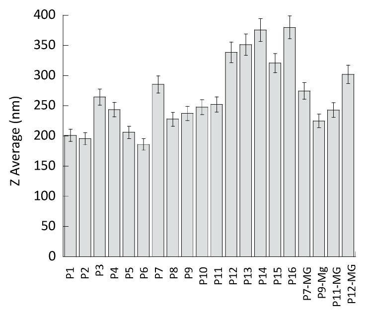
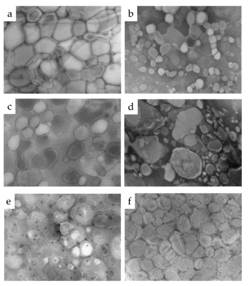
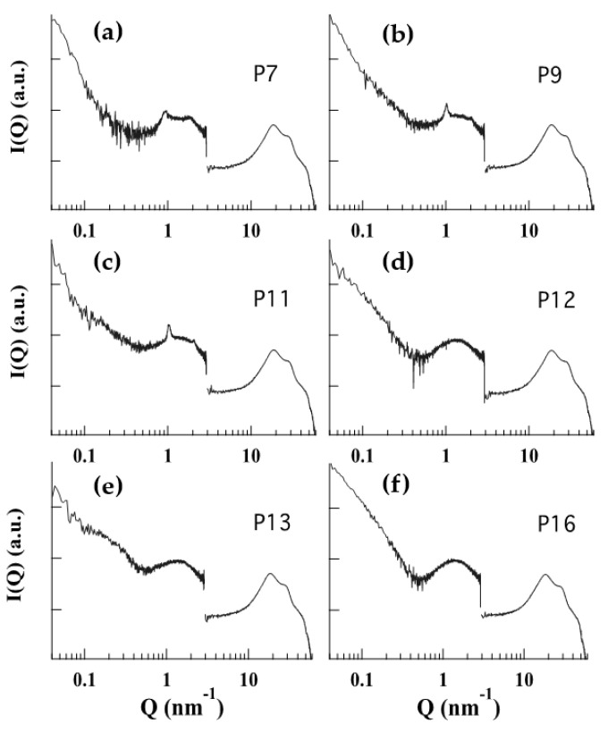
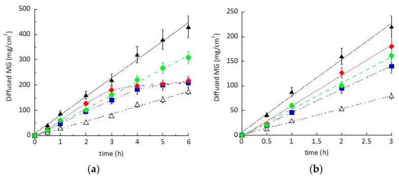
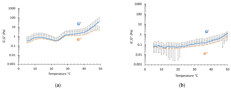
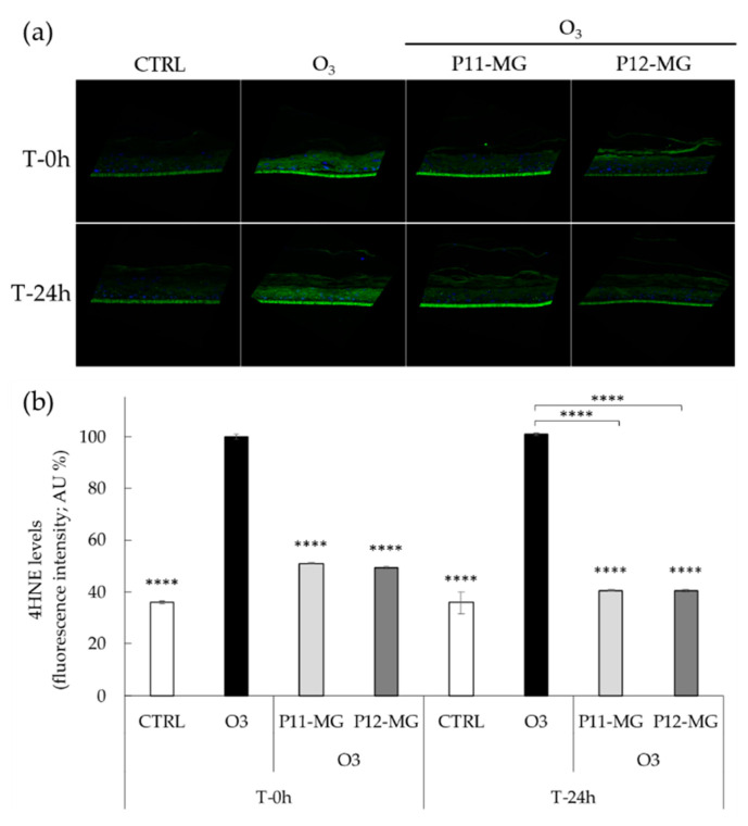
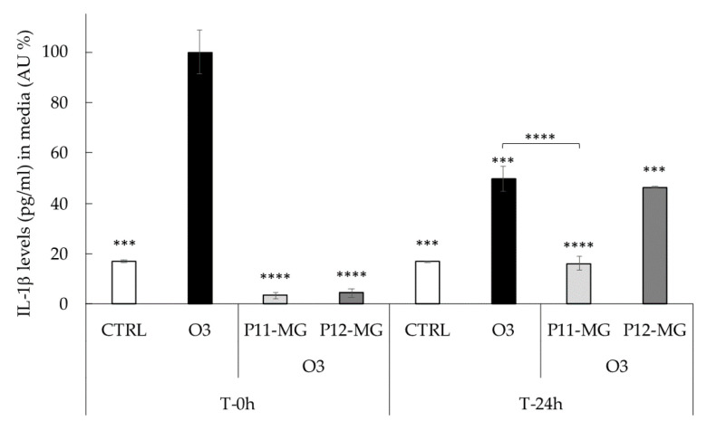
Human skin is dramatically exposed to toxic pollutants such as ozone. To counteract the skin disorders induced by the air pollution, natural antioxidants such as mangiferin could be employed. A formulative study for the development of vesicular systems for mangiferin based on phosphatidylcholine and the block copolymer pluronic is described. Plurethosomes were designed for mangiferin transdermal administration and compared to ethosome and transethosome. Particularly, the effect of vesicle composition was investigated on size distribution, inner and outer morphology by photon correlation spectroscopy, small angle X-ray diffraction, and transmission electron microscopy. The potential of selected formulations as vehicles for mangiferin was studied, evaluating encapsulation efficiency and in vitro diffusion parameters by Franz cells. The mangiferin antioxidant capacity was verified by the 2,2-diphenyl-1-picrylhydrazyl assay. Vesicle size spanned between 200 and 550 nm, being influenced by phosphatidylcholine concentration and by the presence of polysorbate or pluronic. The vesicle supramolecular structure was multilamellar in the case of ethosome or plurethosome and unilamellar in the case of transethosome. A linear diffusion of mangiferin in the case of ethosome and transethosomes and a biphasic profile in the case of plurethosomes indicated the capability of multilamellar vesicles to retain the drug more efficaciously than the unilamellar ones. The antioxidant and anti-inflammatory potential effect of mangiferin against pollutants was evaluated on 3D human skin models exposed to O3. The protective effect exerted by plurethosomes and transethosomes suggests their possible application to enhance the cutaneous antioxidant defense status.

Keywords: antioxidant; in vitro diffusion; mangiferin; phosphatidylcholine; poloxamer.


Conflict of interest statement

The authors declare no conflict of interest. The funders had no role in the design of the study; in the collection, analyses, or interpretation of data; in the writing of the manuscript, or in the decision to publish the results.

Figures

Figure 1
Figure 1
Mean diameter of the indicated formulations. Diameters were measured by PCS and expressed as Z average (nm). Data are the mean of three determinations on different samples ± s.d.
Figure 2
Figure 2
Transmission electron microscopy images (TEM) of P7 (a), P9 (b), P11 (c), P12 (d), P13 (e), and P16 (f). Bar corresponds to 200 nm (a,c,d) or 500 nm (b,e,f).
Figure 3
Figure 3
SAXS/WAXS profiles observed at 35 °C. P7 (a), P9 (b), P11 (c), P12 (d), P13 (e), and P16 (f).
Figure 4
Figure 4
MG diffusion kinetics from Sol-MG (closed triangles), P7-MG (open triangles), P9-MG (green circles), P11-MG (red rhombuses), and P12-MG (blue squares), as determined by Franz cell. Experiments were conducted for 6 h (a), with linear profiles within 3 h (b). Data are the mean of 6 independent experiments ± s.d.
Figure 5
Figure 5
Evolution of elastic (G′, blue) and viscous (G″, orange) moduli for P11-MG (a) and P12-MG (b).
Figure 6
Figure 6
(a) Representative images of immunofluorescence staining for 4HNE protein adducts (green) and DAPI (blue) at 40× magnification on 3D skin models treated with P11-MG and P12-MG and exposed to O3 (b) quantification of immunofluorescence staining of 4HNE protein adducts 0 h and 24 h after O3 exposure. Data were normalized with respect to the O3-exposed sample at 0 h and expressed as arbitrary units (%) ± SD. **** p ≤ 0.0001 vs. O3 at T-0 h. The autofluorescence signal of the 3D skin model inserts was not considered in the quantification analysis.
Figure 7
Figure 7
Levels of IL-1β (pg/mL) in the media of 3D skin models treated with P11-MG and P12-MG analyzed using an IL-1β ELISA kit 0 or 24 h after exposure to O3 0.4 ppm for 4 h. Data were normalized with respect to the O3 sample at 0 h and expressed as arbitrary units (%) ± SD. Data are the results of the averages of at least three different experiments. *** p ≤ 0.001, **** p ≤ 0.0001 vs. O3 at T 0 h.

Similar articles

Cited by

References

    1. Bălă G.-P., Râjnoveanu R.-M., Tudorache E., Motișan R., Oancea C. Air pollution exposure—The (in)visible risk factor for respiratory diseases. Environ. Sci. Pollut. Res. 2021;28:19615–19628. doi: 10.1007/s11356-021-13208-x. - DOI - PMC - PubMed
    1. Lim C.C., Hayes R.B., Ahn J., Shao Y., Silverman D.T., Jones R.R., Garcia C., Bell M.L., Thurston G.D. Long-Term Exposure to Ozone and Cause-Specific Mortality Risk in the United States. Am. J. Respir. Crit. Care Med. 2019;200:1022–1031. doi: 10.1164/rccm.201806-1161OC. - DOI - PMC - PubMed
    1. Ferrara F., Pambianchi E., Woodby B., Messano N., Therrien J.-P., Pecorelli A., Canella R., Valacchi G. Evaluating the effect of ozone in UV induced skin damage. Toxicol. Lett. 2021;338:40–50. doi: 10.1016/j.toxlet.2020.11.023. - DOI - PubMed
    1. Song H., Tan W., Zhang X. Ozone Induces Inflammation in Bronchial Epithelial Cells. J. Asthma. 2011;48:79–83. doi: 10.3109/02770903.2010.529224. - DOI - PubMed
    1. He C., Cheng J., Zhang X., Douthwaite M., Pattisson S., Hao Z. Recent Advances in the Catalytic Oxidation of Volatile Organic Compounds: A Review Based on Pollutant Sorts and Sources. Chem. Rev. 2019;119:4471–4568. doi: 10.1021/acs.chemrev.8b00408. - DOI - PubMed

LinkOut - more resources