. 2021 Jul 23;13(8):1130.
doi: 10.3390/pharmaceutics13081130.

Pheophorbide A and Paclitaxel Bioresponsive Nanoparticles as Double-Punch Platform for Cancer Therapy

Affiliations

Pheophorbide A and Paclitaxel Bioresponsive Nanoparticles as Double-Punch Platform for Cancer Therapy

Francesca Moret et al. Pharmaceutics. .

Abstract

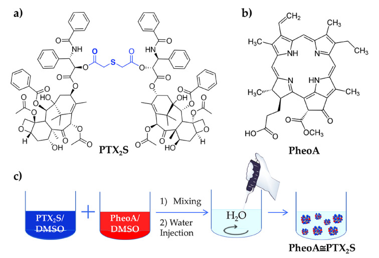
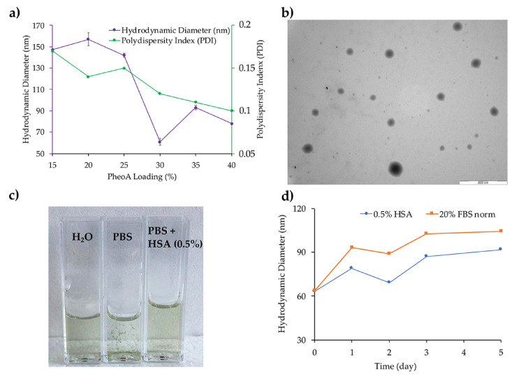
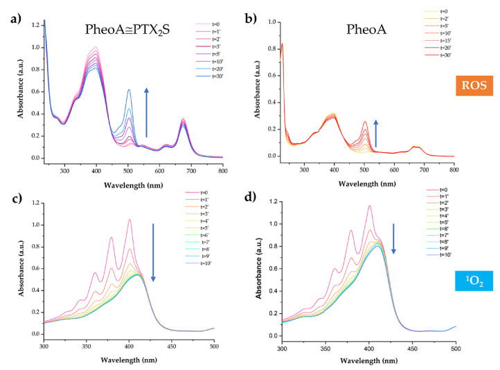
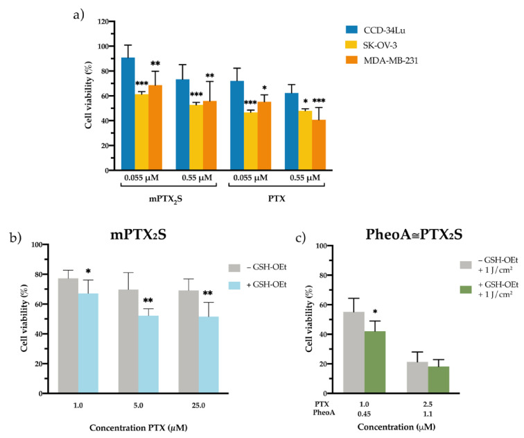
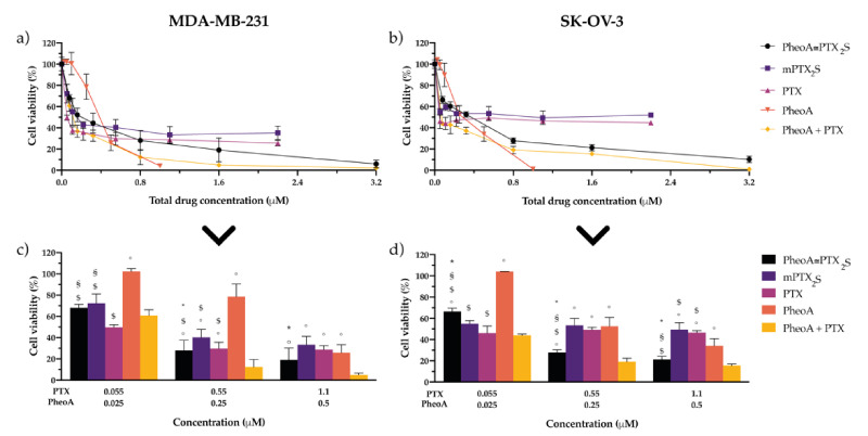
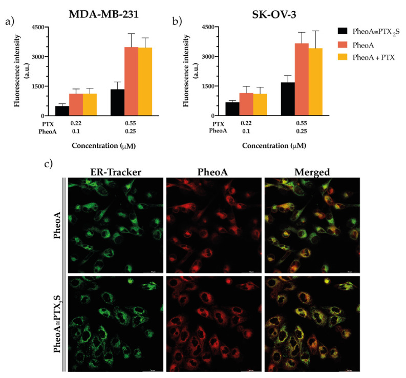
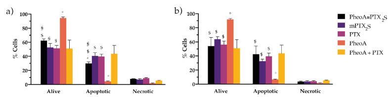
Cancer therapy is still a challenging issue. To address this, the combination of anticancer drugs with other therapeutic modalities, such as light-triggered therapies, has emerged as a promising approach, primarily when both active ingredients are provided within a single nanosystem. Herein, we describe the unprecedented preparation of tumor microenvironment (TME) responsive nanoparticles exclusively composed of a paclitaxel (PTX) prodrug and the photosensitizer pheophorbide A (PheoA), e.g., PheoA≅PTX2S. This system aimed to achieve both the TME-triggered and controlled release of PTX and the synergistic/additive effect by PheoA-mediated photodynamic therapy. PheoA≅PTX2S were produced in a simple one-pot process, exhibiting excellent reproducibility, stability, and the ability to load up to 100% PTX and 40% of PheoA. Exposure of PheoA≅PTX2S nanoparticles to TME-mimicked environment provided fast disassembly compared to normal conditions, leading to PTX and PheoA release and consequently elevated cytotoxicity. Our data indicate that PheoA incorporation into nanoparticles prevents its aggregation, thus providing a greater extent of ROS and singlet oxygen production. Importantly, in SK-OV-3 cells, PheoA≅PTX2S allowed a 30-fold PTX dose reduction and a 3-fold dose reduction of PheoA. Our data confirm that prodrug-based nanocarriers represent valuable and sustainable drug delivery systems, possibly reducing toxicity and expediting preclinical and clinical translation.

Keywords: nanoparticles; paclitaxel; pheophorbide A; photodynamic therapy; prodrug; tumor microenvironment.


Conflict of interest statement

The authors declare no conflict of interest. The funders had no role in the design of the study; in the collection, analyses, or interpretation of data; in the writing of the manuscript, or in the decision to publish the results.

Figures

Figure 1
Figure 1
(a) Chemical structure of PTX2S; (b) Chemical structure of PheoA; (c) Schematic representation of PheoA≅PTX2S preparation by the nanoprecipitation method.
Figure 2
Figure 2
(a) PheoA≅PTX2S optimization as a function of PheoA loading; (b) TEM analysis of PheoA≅PTX2S (scale bar 500 nm; (c) Comparison between PheoA≅PTX2S solution in water, PBS and PBS + 0.5% HSA; (d) PheoA≅PTX2S stability trend in PBS + 0.5% HSA and PBS + 20% FBS.
Figure 3
Figure 3
PheoA≅PTX2S ability to produce ROS and 1O2 upon light irradiation. Absorption spectra of 2′,7′-dichlorofluorescein (DCF) measured at different irradiation times in the presence of (a) PheoA≅PTX2S and (b) free PheoA. 1O2 analysis performed at different irradiation times of a solution of (c) DMA + PheoA≅PTX2S and (d) DMA + PheoA.
Figure 4
Figure 4
In vitro cytotoxicity of nanoparticles in different microenvironments. (a) Cell viability measured in normal fibroblasts (CCD-34-Lu) and cancer cells (breast MDA-MB-231 and ovarian SK-OV-3) incubated for 24 h with increasing concentrations of mPTX2S or PTX delivered in the standard solvent and measured 24 h post cell-release in drug-free medium. * p < 0.05; ** p < 0.01; *** p < 0.001 significantly different from CCD-34Lu (ANOVA One-way with Bonferroni’s correction). (b) Cytotoxicity induced by mPTX2S measured in MDA-MB-231 cells pre-treated or not with GSH-OEt (90 min) before nanoparticles incubation (5 h), to further increase the reductive microenvironment in vitro. (c) Cytotoxicity measured in MDA-MB-231 cells (pre-treated or not with GSH-OEt) incubated with PheoA≅PTX2S and irradiated with red light (1 J/cm2). * p < 0.05; ** p < 0.01 significantly different from-GSH-OEt (Student’s t-test). All data in the figure are expressed as mean ± S.D. of at least two independent experiments carried out in triplicate.
Figure 5
Figure 5
In vitro combination therapy. Dose-response curves of (a) MDA-MB-231 or (b) SK-OV-3 cells incubated for 24 h with single drugs or their combination delivered free or in nanoparticles and irradiated with 1 J/cm2 of light; after an additional 24 h in drug-free medium, cell viability was measured by MTS assay. Total drug concentration is referred to PTX + PheoA concentration. Data are expressed as mean percentage ±SD of at least three independent experiments, carried out in triplicate. To better compare the cytotoxic effects of the different drug formulations, some concentrations have been extrapolated: panel (c) for MDA-MB-231, and panel (d) for SK-OV-3 cells. Statistical significance was calculated applying the ANOVA Two-way with Bonferroni’s correction: * significantly different from mPTX2S; § significantly different from PTX; $ significantly different from PheoA; ° significantly different from PheoA + PTX.
Figure 6
Figure 6
In vitro uptake and intracellular localization studies. Flow cytometry measurements of the intracellular uptake of PheoA delivered in the different formulations in (a) MDA-MB-231 and (b) SK-OV-3 cells exposed to the treatments for 4 h. Data are expressed as mean percentage ± SD of at least three independent experiments, carried out in triplicate. (c) Confocal microscopy images of MDA-MB-231 cells showing the co-localization between the red fluorescence of PheoA (delivered in the standard solvent or loaded in PheoA≅PTX2S) and the green fluorescence of ER-Tracker used as a specific probe for endoplasmic reticulum. Scale bars: 40 µm.
Figure 7
Figure 7
In vitro studies on cell death mechanism. MDA-MB-231 (a) and SK-OV-3 (b) cells incubated with the different drug formulations for 24 h, irradiated with 1 J/cm2 of red light and stained with the Annexin V/PI kit 12 h post-irradiation. Data are expressed as mean percentage ±SD of at least two independent experiments, carried out in triplicate. Statistical significance was calculated applying the ANOVA Two-way with Bonferroni’s correction: § significantly different from PTX; $ significantly different from PheoA; ° significantly different from PheoA + PTX.

References

    1. Misra R., Acharya S., Sahoo S.K. Cancer nanotechnology: Application of nanotechnology in cancer therapy. Drug Discov. Today. 2010;15:842–850. doi: 10.1016/j.drudis.2010.08.006. - DOI - PubMed
    1. Bayat Mokhtari R., Homayouni T.S., Baluch N., Morgatskaya E., Kumar S., Das B., Yeger H. Combination therapy in combating cancer. Oncotarget. 2017;8:38022–38043. doi: 10.18632/oncotarget.16723. - DOI - PMC - PubMed
    1. Zuluaga M.-F., Lange N. Combination of Photodynamic Therapy with Anti-Cancer Agents. Curr. Med. Chem. 2008;15:1655–1673. doi: 10.2174/092986708784872401. - DOI - PubMed
    1. Cao J., Chen Z., Chi J., Sun Y., Sun Y. Recent progress in synergistic chemotherapy and phototherapy by targeted drug delivery systems for cancer treatment. Artif. Cells Nanomed. Biotechnol. 2018;46:817–830. doi: 10.1080/21691401.2018.1436553. - DOI - PubMed
    1. Brown S.B., Brown E.A., Walker I. The present and future role of photodynamic therapy in cancer treatment. Lancet Oncol. 2004;5:497–508. doi: 10.1016/S1470-2045(04)01529-3. - DOI - PubMed

LinkOut - more resources