Review
. 2021 Aug 16;13(8):1262.
doi: 10.3390/pharmaceutics13081262.

Bioinspired Magnetic Nanochains for Medicine

Affiliations
Review

Bioinspired Magnetic Nanochains for Medicine

Slavko Kralj et al. Pharmaceutics. .

Abstract

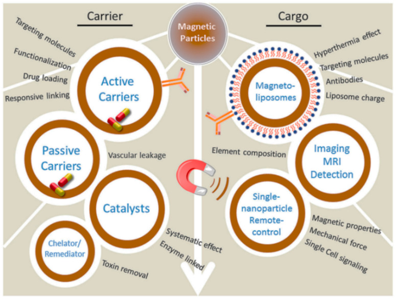
Superparamagnetic iron oxide nanoparticles (SPIONs) have been widely used for medicine, both in therapy and diagnosis. Their guided assembly into anisotropic structures, such as nanochains, has recently opened new research avenues; for instance, targeted drug delivery. Interestingly, magnetic nanochains do occur in nature, and they are thought to be involved in the navigation and geographic orientation of a variety of animals and bacteria, although many open questions on their formation and functioning remain. In this review, we will analyze what is known about the natural formation of magnetic nanochains, as well as the synthetic protocols to produce them in the laboratory, to conclude with an overview of medical applications and an outlook on future opportunities in this exciting research field.

Keywords: biocompass; biomineralization; magnetic assembly; magnetic navigation; magnetite; magnetoreception; magnetosome chains; magnetotactic bacteria; single domain particles; superparamagnetic iron oxide nanoparticles.


Conflict of interest statement

The authors declare no conflict of interest.

Figures

Figure 2
Figure 2
Schematic presentation of magnetotactic bacterium with a magnetosome chain. The magnetosome consists of lipid invaginations, each one enclosing a ferrimagnetic nanocrystal. The 1D chain of magnetosomes is decorated by MamA homo-oligomers. Reproduced from [24]. Copyright 2011, with permission from PNAS.
Figure 1
Figure 1
Biomedical applications for magnetic nanoparticles. MRI = magnetic resonance imaging. Reproduced from [2]. CC BY 4.0 license.
Figure 3
Figure 3
Transmission electron microscopy (TEM) images of magnetotactic bacteria with 1D chains of magnetosome nanoparticles of different morphologies. (a) Magnetovibrio blakemorei strain MV-1 where elongated prismatic nanocrystals can be found, (b) Desulfovibrio magneticus strain RS-1 which produces bullet-shaped nanoparticles, and (c) TEM images of magnetosome chain from a lysed cell of Magnetospirillum magneticum, strain AMB-1. Each cuboctahedral magnetite nanocrystal is surrounded by the phospholipid magnetosome membrane, which often remains stable even after cell lysis. Adapted from [17], Copyright 2013, with permission from Elsevier.
Figure 4
Figure 4
Proteins that are potentially involved in the different phases of magnetosome formation and assembly. (A) Magnetosome membrane formation; in particular, MamY (blue) could be used to shape and close the vesicle, and to sort further proteins. (B) Crystallization of magnetite, with MamO in yellow. (C) Particle size control. (D) Chain assembly, with MamJ (green) to anchor proteins, MamK filament proteins in orange, and potentially MamA. Reproduced with permission from [48], Copyright 2015, with permission from Wiley.
Figure 5
Figure 5
Cellular organization of bacterial magnetosomes. Reprinted from [70] copyright 2006, with permission from Elsevier.
Figure 6
Figure 6
TEM images and corresponding schematic illustrations of synthesized magnetically guidable nanoparticle clusters. (a) Composite magnetic nanoparticle cluster composed of superparamagnetic maghemite nanoparticles and biocompatible polymer [85] (b) One-pot solvothermal synthesis of clusters composed of many superparamagnetic magnetite nanoparticles. Adapted with permission from [84] Copyright 2007, with permission from Wiley. (c) Emulsion/evaporation-based clustering of hydrophobic maghemite nanoparticles following silica coating. Reprinted from [29], Copyright 2014, with permission from Elsevier. (d) Chemical cross-linking method of differently functionalized superparamagnetic maghemite nanoparticles forming synthesized magnetically guidable nanoparticle cluster, adapted from [42]. CC BY 4.0 license.
Figure 7
Figure 7
Schematic illustration and corresponding TEM images of key steps in the magnetic nanochain formation. (a) Alignment of superparamagnetic maghemite nanoparticle clusters in dynamic magnetic field. (b) Nanochains’ building blocks consisting of silica-coated nanoparticle clusters. Inset: the resulting suspension is a magnetically tunable photonic crystalline liquid resulting in a structural color when the suspension is exposed to a weak external magnetic field. (c) Rigid magnetic nanochains coated with fixating silica shell. (d) Short nanochains composed of ca. 6 clusters (length ca. 400–700 nm). (e) Magnetic nanochains composed of ca. 14 clusters (length ca. < 1 µm). (f) Short inter-particle distance between adjacent particles in the nanochain by choosing building blocks with thin primary silica shell of 5 nm. (g) Large inter-particle distance between adjacent particles in the nanochain by choosing building blocks with thick primary silica shell of 20 nm. Reprinted with permission from [28], Copyright © 2021, with pemission from American Chemical Society.
Figure 8
Figure 8
Schematic illustration of the fabrication process of short peapod-like magnetic nanochains. (a) Schematic synthesis protocol and (b) TEM image of the resulting nanochains. Reprinted with permission from [106], Copyright © 2021, with pemission from American Chemical Society.
Figure 9
Figure 9
(ac) Schematic illustration of a method for fabricating striped patterns of magnetic nanoparticles. (a) Template consisting of elongated ridges and gaps, and a flat target substrate. (b) After suspension deposition, water is evaporated and the film is divided in the sections along the ridges of the template. (c) 1D arrangement of the nanoparticles. Reproduced with permission from [107], Copyright 2016, with permission from Wiley. (d,e) Colloidal approach for the magnetic structuring in suspension. Reproduced with permission from [28], Copyright © 2021, with pemission from American Chemical Society. (d) Low-magnification TEM image of anisotropic magnetic nanobundles composed of superparamagnetic nanoparticle clusters, and (e) TEM image of one magnetic nanobundle.
Figure 10
Figure 10
Reaction scheme of the controlled assembly of magnetic nanochains using solid-phase chemistry. (a) Precise control over the chemical functionality of amines and thiols on the surface of individual magnetic nanoparticles. (b) TEM image of fabricated nanochains, each one composed of three units of nanoparticles. (c) Schematic illustration of a multi-step process to produce nanochains with a terminally bonded liposome drug carrier. Reproduced from [110], Copyright 2013, with permission from Elsevier.
Figure 11
Figure 11
Schematic illustration of ferrimagnetic nanochain assembly, their surface modification, transfection of mesenchymal stem cells, and in vivo application to trigger post-stroke recovery in a mouse model. Reproduced with permission from [26], Copyright 2019, with permission from Wiley.
Figure 12
Figure 12
Future application of multifunctional short nanochains carrying drug molecules. (a) Schematic representation of short nanochain with radially aligned pores on the surface, whose design and production has recently been described [39]. (b) Schematic representation of drug-loaded nanochains. (c) Drug release can be triggered through exposure to a rotating magnetic field (RMF). Reproduced from [39] under a Creative Commons license.
Figure 13
Figure 13
Schematic presentation of therapeutic anticancer approach based on nanochains following three steps. (1) Short nanochain loaded with drug circulates in the blood. (2) Nanochains’ target micrometastases. (3) Radiofrequency-triggered drug release from the nanochains. Reproduced from [110] Copyright 2013, with permission from Elsevier.

References

    1. Huang C., Chen X., Xue Z., Wang T. Effect of structure: A new insight into nanoparticle assemblies from inanimate to animate. Sci. Adv. 2020;6:eaba1321. doi: 10.1126/sciadv.aba1321. - DOI - PMC - PubMed
    1. Kudr J., Haddad Y., Richtera L., Heger Z., Cernak M., Adam V., Zitka O. Magnetic Nanoparticles: From Design and Synthesis to Real World Applications. Nanomaterials. 2017;7:243. doi: 10.3390/nano7090243. - DOI - PMC - PubMed
    1. Zhang T., Xu Q., Huang T., Ling D., Gao J. New Insights into Biocompatible Iron Oxide Nanoparticles: A Potential Booster of Gene Delivery to Stem Cells. Small. 2020;16:2001588. doi: 10.1002/smll.202001588. - DOI - PubMed
    1. Farzin A., Etesami S.A., Quint J., Memic A., Tamayol A. Magnetic Nanoparticles in Cancer Therapy and Diagnosis. Adv. Healthc. Mater. 2020;9:1901058. doi: 10.1002/adhm.201901058. - DOI - PMC - PubMed
    1. Xie W., Guo Z., Gao F., Gao Q., Wang D., Liaw B., Cai Q., Sun X., Wang X., Zhao L. Shape-, size- and structure-controlled synthesis and biocompatibility of iron oxide nanoparticles for magnetic theranostics. Theranostics. 2018;8:3284–3307. doi: 10.7150/thno.25220. - DOI - PMC - PubMed

LinkOut - more resources