. 2021 Aug 17;13(8):1277.
doi: 10.3390/pharmaceutics13081277.

Effective Osteogenic Priming of Mesenchymal Stem Cells through LNA-ASOs-Mediated Sfrp1 Gene Silencing

Affiliations

Effective Osteogenic Priming of Mesenchymal Stem Cells through LNA-ASOs-Mediated Sfrp1 Gene Silencing

Daniel García-Sánchez et al. Pharmaceutics. .

Abstract

Mesenchymal stem cell (MSC) transplantation has emerged as a promising approach for bone regeneration. Importantly, the beneficial effects of MSCs can be improved by modulating the expression levels of specific genes to stimulate MSC osteogenic differentiation. We have previously shown that Smurf1 silencing by using Locked Nucleic Acid-Antisense Oligonucleotides, in combination with a scaffold that sustainably releases low doses of BMP-2, was able to increase the osteogenic potential of MSCs in the presence of BMP-2 doses significantly smaller than those currently used in the clinic. This would potentially allow an important reduction in this protein in MSs-based treatments, and thus of the side effects linked to its administration. We have further improved this system by specifically targeting the Wnt pathway modulator Sfrp1. This approach not only increases MSC bone regeneration efficiency, but is also able to induce osteogenic differentiation in osteoporotic human MSCs, bypassing the need for BMP-2 induction, underscoring the regenerative potential of this system. Achieving successful osteogenesis with the sole use of LNA-ASOs, without the need of administering pro-osteogenic factors such as BMP-2, would not only reduce the cost of treatments, but would also open the possibility of targeting these LNA-ASOs specifically to MSCs in the bone marrow, allowing us to treat systemic bone loss such as that associated with osteoporosis.

Keywords: BMP; GapmeR; LNA-ASO; Sfrp1; bone regeneration; mesenchymal stem cells; osteogenesis.


Conflict of interest statement

The authors declare no conflict of interest.

Figures

Figure 1
Figure 1
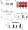
Analysis of proliferation, apoptosis, and migration of MSCs transfected with GapmeRs specific for the silencing of key osteogenic inhibitors. (a) Silencing of the different targeted genes in C3H10T1/2 MSCs using specific GapmeRs 48 h after transfection. Ctrl stands for Control GapmeR (n = 7). Error bars represent standard error of the mean values. (b) Representative picture of the immunofluorescence study of Ki67 expression in cells transfected with the different GapmeRs proliferating in normal culture media at different timepoints. Graph represents the increment of fluorescent cells referred to the previous day in each of the conditions tested. (c) Flow cytometry analysis of apoptosis using Annexin V and 7AAD. Average values of three different flow cytometry analysis are shown. Cells were stained with Annexin V (AnnV) and propidium iodide (PI) to detect early apoptosis (Ann+), late apoptosis (Ann/PI+), or dead cells (PI+). Percentage of positive cells for each population was normalized to cells transfected with the control GapmeR (Ctrl). Flow cytometry profile is shown to define populations. (d) Wound healing assay. Representative images of cells underperforming in the assay are shown from three independent experiments. Areas lacking cells are outlined. The graph shows the average values and the standard error of at least six different points along the wound measured for each of the samples from a representative experiment.
Figure 1
Figure 1
Analysis of proliferation, apoptosis, and migration of MSCs transfected with GapmeRs specific for the silencing of key osteogenic inhibitors. (a) Silencing of the different targeted genes in C3H10T1/2 MSCs using specific GapmeRs 48 h after transfection. Ctrl stands for Control GapmeR (n = 7). Error bars represent standard error of the mean values. (b) Representative picture of the immunofluorescence study of Ki67 expression in cells transfected with the different GapmeRs proliferating in normal culture media at different timepoints. Graph represents the increment of fluorescent cells referred to the previous day in each of the conditions tested. (c) Flow cytometry analysis of apoptosis using Annexin V and 7AAD. Average values of three different flow cytometry analysis are shown. Cells were stained with Annexin V (AnnV) and propidium iodide (PI) to detect early apoptosis (Ann+), late apoptosis (Ann/PI+), or dead cells (PI+). Percentage of positive cells for each population was normalized to cells transfected with the control GapmeR (Ctrl). Flow cytometry profile is shown to define populations. (d) Wound healing assay. Representative images of cells underperforming in the assay are shown from three independent experiments. Areas lacking cells are outlined. The graph shows the average values and the standard error of at least six different points along the wound measured for each of the samples from a representative experiment.
Figure 2
Figure 2
Effect of Cby1 and Sfrp1 silencing on canonical and non-canonical Wnt pathways. Each experiment was performed in duplicates. A representative experiment is shown. (a) Western blot of JNK and PKC phosphorylation used as markers of the Wnt/PCP and the Wnt/PKC pathway activation. Forty-eight hours after transfection with the different GapmeRs, osteogenic media was added for an additional 48 h before whole protein extracts were obtained. β-actin was used as a loading control. (b) Presence of the nuclear and cytoplasmic β-catenin was quantified in nuclear and cytoplasmic cell extracts. Lamin A/C and Gapdh were used for normalization of the nuclear and cytoplasmic signals, respectively.
Figure 3
Figure 3
Osteogenic differentiation capacity of MSCs after the silencing of specific osteogenic inhibitors and their combinations. Sm1 stands for Smurf1 in the GapmeR combinations. All analyses were performed on transfected cells that had undergone osteogenic differentiation for 11 days. All graphs represent average values of independently performed experiments. Error bars represent standard error of the mean values. For analysis performed on cells transfected with a single GapmeR (n = 7) and for those performed with cells transfected with GapmeR combinations (n = 5). (a) Expression of the osteoblastic differentiation driver Runx2 and osteogenic markers Alpl and Bglap. (b) Alizarin Red staining of cells transfected with GapmeRs or GapmeRs combinations growing in osteogenic media. The picture shows a representative experiment performed at day 11 of differentiation. (c) Graphs shows quantification of the Alizarin Red staining at 405 nm. (d) Graph shows alkaline phosphatase activity measured in the different conditions. (* p ≤ 0.05; ** p ≤ 0.005).
Figure 4
Figure 4
In vivo analysis of the pro-osteogenic effect of the silencing of key osteogenic inhibitors in MSCs. Pictures show histological analysis of sections obtained from representative decalcified implants. All implants were isolated from the same individual. (a) Masson–Goldner staining of subdermal implants in mice, showing mineralized matrix. (b) Representative images of Masson–Goldner staining at higher magnification. White arrowheads indicate osteocyte-like cells surrounded by lacunae and immersed in the mineralized matrix. (c) Histological sections observed under green, fluorescent light show the typical autofluorescence of collagen fibers in the different samples analyzed. (d) Bar graph shows quantification of fluorescence signal intensity in the different histological samples performed using ImageJ. Sm1 stands for Smurf1. One implant of each type was quantified. Values correspond to the average of at least six different areas measured in each of the implants. Error bars represent standard error of the mean values. Sm1 stands for Smurf1 in the Gapmer combinations. (e) Immunohistochemistry of vascular marker CD34. Microvascular density was determined by counts of CD34-positive staining blood vessels. Sm1 stands for Smurf1 in the Gapmer combinations. The graph shows the average values and the standard error of at least five different areas analyzed for each sample. Microscopic appearance of CD34-positive staining blood vessels is shown on the right. Stained microvessels are outlined (** p ≤ 0.005; *** p ≤ 0.0005).
Figure 5
Figure 5
Effect of SFRP1 silencing on the osteogenic potential of human MSCs from osteoporotic patients. All analyses were performed at 21 days of osteogenic differentiation. (a) Semi-quantitative PCRs showing the relative expression levels of different osteogenic markers (RUNXx2, ALPL, and BGLAP) in hMSCs from osteoporotic patients (MSC-OP) transfected with a control GapmeR (Ctrl) and a GapmeR for the silencing of SFRP1 expression. The expression of the analyzed markers in the presence of 10 ng/mL of BMP-2 was used as a reference of the effectiveness of SFRP1 silencing. Values reflect averages of five different samples (n = 5). Technical triplicates were also performed with each individual sample. (b) Graph showing the ALPL activity in MSC-OP under the different experimental conditions analyzed. Graph shows average values of five independent samples. (c) Osteogenic differentiation of the same hMSCs-OP transfected with the SFRP1 GapmeR or differentiated in the presence of 10 ng/mL BMP-2 as revealed by Alizarin Red staining. Results from one representative sample are shown in the pictures. Bar graph represents quantification of the staining. In all graphs, bars represent standard error of the mean values. p-values are indicated.

Similar articles

Cited by

References

    1. Zigdon-Giladi H., Rudich U., Michaeli-Geller G., Evron A. Recent advances in bone regeneration using adult stem cells. World J. Stem Cells. 2015;7:630–640. doi: 10.4252/wjsc.v7.i3.630. - DOI - PMC - PubMed
    1. He X., Dziak R., Yuan X., Mao K., Genco R., Swihart M., Sarkar D., Li C., Wang C., Lu L., et al. BMP2 genetically engineered MSCs and EPCs promote vascularized bone regeneration in rat critical-sized calvarial bone defects. PLoS ONE. 2013;8:e60473. doi: 10.1371/journal.pone.0060473. - DOI - PMC - PubMed
    1. Fierro F.A., Kalomoiris S., Sondergaard C.S., Nolta J.A. Effects on proliferation and differentiation of multipotent bone marrow stromal cells engineered to express growth factors for combined cell and gene therapy. Stem Cells. 2011;29:1727–1737. doi: 10.1002/stem.720. - DOI - PMC - PubMed
    1. Kim D., Cho S.W., Her S.J., Yang J.Y., Kim S.W., Kim S.Y., Shin C.S. Retrovirus-mediated gene transfer of receptor activator of nuclear factor-kappaB-Fc prevents bone loss in ovariectomized mice. Stem Cells. 2006;24:1798–1805. doi: 10.1634/stemcells.2005-0480. - DOI - PubMed
    1. Rodriguez-Evora M., Garcia-Pizarro E., del Rosario C., Perez-Lopez J., Reyes R., Delgado A., Rodriguez-Rey J.C., Evora C. Smurf1 knocked-down, mesenchymal stem cells and BMP-2 in an electrospun system for bone regeneration. Biomacromolecules. 2014;15:1311–1322. doi: 10.1021/bm401854d. - DOI - PubMed

LinkOut - more resources