. 2021 Jul 23;13(8):1433.
doi: 10.3390/v13081433.

3-Indoleacetonitrile Is Highly Effective in Treating Influenza A Virus Infection In Vitro and In Vivo

Affiliations

3-Indoleacetonitrile Is Highly Effective in Treating Influenza A Virus Infection In Vitro and In Vivo

Xuejin Zhao et al. Viruses. .

Abstract

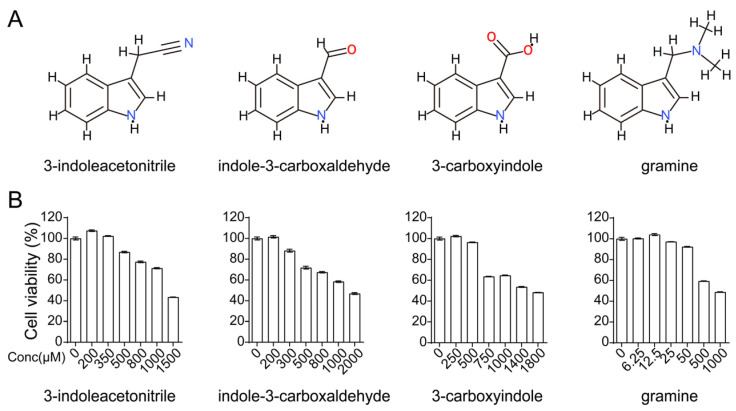
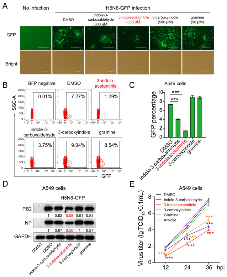
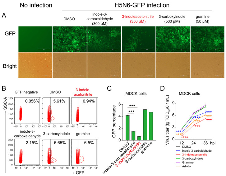
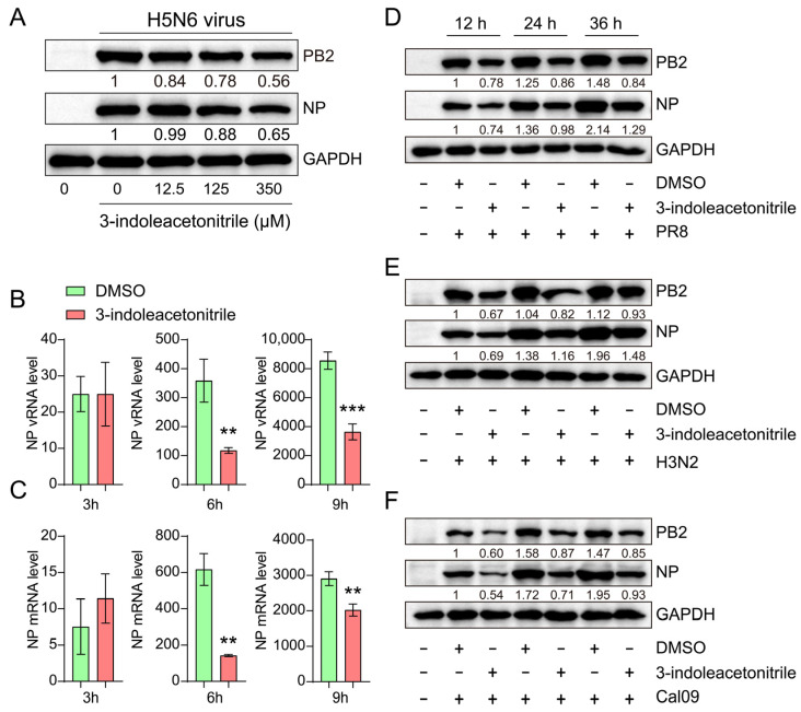
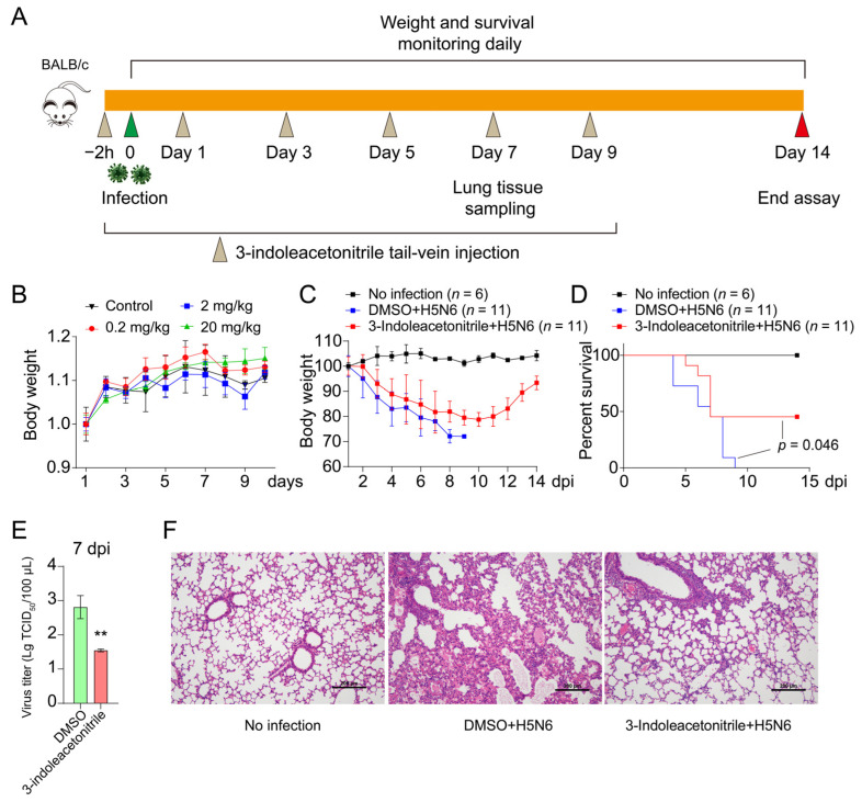
Influenza A viruses are serious zoonotic pathogens that continuously cause pandemics in several animal hosts, including birds, pigs, and humans. Indole derivatives containing an indole core framework have been extensively studied and developed to prevent and/or treat viral infection. This study evaluated the anti-influenza activity of several indole derivatives, including 3-indoleacetonitrile, indole-3-carboxaldehyde, 3-carboxyindole, and gramine, in A549 and MDCK cells. Among these compounds, 3-indoleacetonitrile exerts profound antiviral activity against a broad spectrum of influenza A viruses, as tested in A549 cells. Importantly, in a mouse model, 3-indoleacetonitrile with a non-toxic concentration of 20 mg/kg effectively reduced the mortality and weight loss, diminished lung virus titers, and alleviated lung lesions of mice lethally challenged with A/duck/Hubei/WH18/2015 H5N6 and A/Puerto Rico/8/1934 H1N1 influenza A viruses. The antiviral properties enable the potential use of 3-indoleacetonitrile for the treatment of IAV infection.

Keywords: 3-indoleacetonitrile; antiviral; indole derivatives; indole-3-carboxaldehyde; influenza A virus.


Conflict of interest statement

The authors declare no conflict of interest.

Figures

Figure 1
Figure 1
Cytotoxicity of indole derivatives in A549 cells. (A) Molecular structure of 3-indoleacetonitrile, indole-3-carboxaldehyde, 3-carboxyindole, and gramine. (B) A549 cells were treated with 3-indoleacetonitrile, indole-3-carboxaldehyde, 3-carboxyindole, or gramine using the indicated concentrations for 24 h. Then, the cell viability was measured by CCK-8.
Figure 2
Figure 2
The effect of indole derivatives on H5N6-GFP virus replication. The A549 cells were infected with the H5N6-GFP virus at an MOI of 0.005, followed by treatment with 3-indoleacetonitrile, indole-3-carboxaldehyde, 3-carboxyindole, and gramine at indicated concentrations for 24 h. After that, the GFP intensity was acquired using fluorescence microscopy (A). The percentage of GFP-positive cells was calculated through flow cytometry (B, a presentative image) (C, data collected from three independent biological experiments). The viral PB2 and NP proteins were analyzed by Western blotting (D). The growth curves of H5N6-GFP virus in supernatants were determined based on three time points of infection as indicated (E), 3-indoleacetonitrile (350 μM); indole-3-carboxaldehyde (300 μM); 3-carboxyindole (500 μM); gramine (50 μM); arbidol (8 μM). *** p < 0.001; calculated from three independent experiments by two-tailed Student’s t-test (C) or two-way ANOVA (E).
Figure 3
Figure 3
The effect of indole derivatives on H5N6-GFP virus replication in MDCK cells. The MDCK cells were infected with the H5N6-GFP virus at an MOI of 0.005, followed by treatment with 3-indoleacetonitrile, indole-3-carboxaldehyde, 3-carboxyindole, and gramine at indicated concentrations for 24 h. After that, the GFP intensity was acquired using fluorescence microscopy (A). The percentage of GFP-positive cells was calculated through flow cytometry (B, a presentative image) (C, data collected from three independent biological experiments). The growth curves were determined by measuring H5N6-GFP virus yield in supernatants at time points of 12, 24, and 36 hpi (D), 3-indoleacetonitrile (350 μM); indole-3-carboxaldehyde (300 μM); 3-carboxyindole (500 μM); gramine (50 μM); arbidol (8 μM). *** p < 0.001; calculated from three independent experiments by two-tailed Student’s t-test (C) or two-way ANOVA (D).
Figure 4
Figure 4
3-indoleacetonitrile inhibits influenza virus replication with a broad spectrum. (A) The protein level of H5N6 NP and PB2 was measured in A549 cells treated with an increased dose of 3-indoleacetonitrile. (B,C) The effect of 3-indoleacetonitrile on H5N6 vRNA (B) and mRNA (C) abundance at 3, 6, and 9 hpi. (DF) Effect of 3-indoleacetonitrile on viral PB2 and NP protein level in A549 cells infected with PR8 (D), H3N2 (E), or Cal09 (F) viruses. ** p < 0.01; *** p < 0.001; calculated from three independent experiments by two-tailed Student’s t-test.
Figure 5
Figure 5
3-indoleacetonitrile restricts H5N6 virus replication in mice. (A) Schematic diagram of the mouse experiment. (B) Female BALB/c mice were treated with 3-indoleacetonitrile (0.2, 2 or 20 mg/kg per day) or 0.5% DMSO via tail-vein injection. Body weights in each group were monitored daily for 10 days post-treatment. (CF) Female BALB/c mice were inoculated with 50 μL of 2 LD50 H5N6 viruses and treated with 20 mg/kg 3-indoleacetonitrile or 0.5% DMSO. In the next 2 weeks, the body weights (C) and mouse survival (D) were recorded daily. p-value was calculated using the log-rank (Mantel–Cox) test. On 7 dpi, the left lung’s viral titers (E) were measured by determining the TCID50, and the right lungs were fixed for H&E staining (F). ** p < 0.01; calculated from three independent experiments by two-tailed Student’s t-test.

Similar articles

Cited by

References

    1. Krammer F., Smith G.J.D., Fouchier R., Peiris J.S.M., Kedzierska K., Doherty P.C., Palese P., Shaw M.L., Treanor J., Webster R.G., et al. Influenza. Nat. Rev. Dis. Primers. 2018;4:3. doi: 10.1038/s41572-018-0002-y. - DOI - PMC - PubMed
    1. Trilla A., Trilla G., Daer C. The 1918 “Spanish Flu” in Spain. Clin. Infect. Dis. 2008;47:668–673. doi: 10.1086/590567. - DOI - PubMed
    1. Jester B.J., Uyeki T.M., Jernigan D.B. Fifty Years of Influenza A(H3N2) Following the Pandemic of 1968. Am. J. Public Health. 2020;110:669–676. doi: 10.2105/AJPH.2019.305557. - DOI - PMC - PubMed
    1. LaRussa P. Pandemic Novel 2009 H1N1 Influenza: What Have We Learned? Semin. Respir. Crit. Care Med. 2011;32:393–399. doi: 10.1055/s-0031-1283279. - DOI - PubMed
    1. Buchy P., Badur S. Who and When to Vaccinate Against Influenza. Int. J. Infect. Dis. 2020;93:375–387. doi: 10.1016/j.ijid.2020.02.040. - DOI - PubMed

Publication types

MeSH terms

LinkOut - more resources