Review
. 2021 Jul 24;13(8):1446.
doi: 10.3390/v13081446.

Morphological Diversity and Dynamics of Dengue Virus Affecting Antigenicity

Affiliations
Review

Morphological Diversity and Dynamics of Dengue Virus Affecting Antigenicity

Guntur Fibriansah et al. Viruses. .

Abstract

The four serotypes of the mature dengue virus can display different morphologies, including the compact spherical, the bumpy spherical and the non-spherical clubshape morphologies. In addition, the maturation process of dengue virus is inefficient and therefore some partially immature dengue virus particles have been observed and they are infectious. All these viral particles have different antigenicity profiles and thus may affect the type of the elicited antibodies during an immune response. Understanding the molecular determinants and environmental conditions (e.g., temperature) in inducing morphological changes in the virus and how potent antibodies interact with these particles is important for designing effective therapeutics or vaccines. Several techniques, including cryoEM, site-directed mutagenesis, hydrogen-deuterium exchange mass spectrometry, time-resolve fluorescence resonance energy transfer, and molecular dynamic simulation, have been performed to investigate the structural changes. This review describes all known morphological variants of DENV discovered thus far, their surface protein dynamics and the key residues or interactions that play important roles in the structural changes.

Keywords: antibody complex; dengue; flavivirus; morphological changes.


Conflict of interest statement

The authors declare no conflict of interest.

Figures

Figure 1
Figure 1
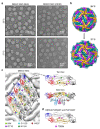
The organization of the structural proteins in DENV. (a) Different arrangement of the E protein or E-prM protein on neutral pH immature (top), low pH immature (center), and neutral pH mature (bottom) particles that are present in the maturation process of DENV. The black triangles indicate an icosahedral asymmetric unit (asu), with 5-, 3- and 2-fold vertices shown. Each asu contains 3 individual E proteins, which are indicated as molecules A, B and C. The E proteins in the neighboring asu are labeled A′, B′, and C′. They are each located near to 5-, 2- and 3-fold vertices and are coloured in gold, dark cyan, and purple, respectively. (b) E and prM proteins in immature DENV exist as a heterodimer, and three E-prM heterodimers form an inverted tripod structure. (Top, right) The E protein ectodomain consists of DI (red), DII (yellow) and DIII (dark blue), which is connected to membrane helices (blue) via three stem helices (cyan). PrM protein consist of pr-peptide (light green), a long loop that contains furin cleavage site, stem helices (pink) and then trans-membrane helices (dark pink). (Bottom, right) Three E-prM heterotrimers form an inverted tripod structure and a C protein dimer bound to the trans membrane helices region of the inverted tripod. The three E-prM heterodimers that form an inverted tripod are colored in gold, dark cyan, and purple. The helices α1–α5 of one of the C protein protomer in a dimer are colored in a gradient of brown shades, whereas the the other protomer is colored in gray. (Bottom, left) Three inverted tripod structure interact to each other to form the building block for the immature virus shell. The three C protein dimers further make a triangular network strengthening the building block structure. (Top, left) One building block interacts with another through the tips of their E–prM heterodimers forming the spiky-looking lattice of the immature virus. Three neighboring building blocks (numbered 1 to 3) are shown with colors whereas the others are in light gray. Building block 1 (indicated in dashed line) has one inverted tripod colored in gold-dark cyan-purple, whereas the other two inverted tripods are in two different shades of gray. Building blocks 2 and 3 are colored in brown and olive green, respectively. (c) An E protein dimer that presence on the mature DENV structure. One E protein protomer in a dimer is colored as in panel b (top, right), whereas the other E protein is colored in gray.
Figure 2
Figure 2
Structural changes in DENV2 strain NGC at 37 °C. (a) CryoEM micrographs of the lab-adapted DENV2 NGC strains with different passage histories in mammalian (BHK21) and mosquito (C6/36) cell lines, when incubated at 29 and 37 °C. (b) The E protein on DENV2 surface undergoes structural rearrangement upon incubation at 37 °C. The organization of E proteins on DENV2 at 29 °C (top) and 37 °C (bottom) is shown. At 37 °C, the E protein mols A-C′ particle remains as a dimer, but it undergoes translation and rotation, whereas the E protein molecules B and B′ move apart from each other, and thus are no longer dimers. Furthermore, the E protein molecule B/B′ is rotated upwards using the fusion loop as a pivot point, resulting in the protusion of DIII. The black triangle represents an icosahedral asymmetric unit with the corresponding 5-, 2-, and 3-fold vertices indicated. (c) DENV2 NGC (BHK21) showed differences in their E protein sequences at 5 positions when compared to DENV2-NGC (C6/36). (Left) The locations of these 5 residue differences are shown on an E protein raft on the virus surface. The residue mutations are shown as spheres. (Right) Top and side view of an E protein dimer showing the locations of the 5 residue differences. Top view of the E protein dimer only shows the ectodomain part, whereas the side view includes the TM regions. Residue 6 (I6M) is indicated by magenta dashed circle. (d) The location of one residue difference on E protein observed between DENV2 strain PVP94/07 and DENV2 strain PVP103/07. DENV2 strain PVP103/07 displayed morphological changes at 37 °C, whereas DENV2 strain PVP94/07 not. The E protein is colored as in Figure 1.
Figure 3
Figure 3
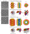
Helical structures of the tail of DENV3-Fab C10 clubSP and ZIKV-Fab C10 catSP. (a) CryoEM micrographs of the lab-adapted DENV3 CH53489 at 4, 29 and 37 °C. DENV3 at 4 °C contains mostly spherical particles, and when incubated at 29 and 37 °C, clubSP particles will form. In addition, at 37 °C, some of the smooth spherical-shaped particles also adopt a bumpy morphology. (b,c) (Top) The cryoEM map of the tail of DENV3-Fab C10 clubSP (b) and ZIKV-Fab C10 catSP (c). The side view (left), central cross-section (center), and top view of the reconstructed helical map are shown. For DENV3-Fab C10 clubSP, the map is colored according to the radii from the helical axis: blue (10–30 Å), green (31–50 Å), yellow (51–90 Å), and red (91–140 Å), whereas for ZIKV-Fab C10 catSP: blue (10–75 Å), green (76–105 Å), yellow (106–140 Å), and red (146–195 Å). These colors correspond to the inner leaflet of the lipid bilayer membrane, the transmembrane regions of the E and M proteins, the ectodomain of the E and M proteins, and Fab C10, respectively. (Bottom) The proposed mechanism of how the E proteins rearrange from the original smooth compact spherical virus structure to form the final helical structure. The DENV3 CH53489-Fab C10 clubSP structure (b) shows that Fab C10 does not lock the DENV3 raft structure. The Fab C10 only locks the E protein dimer and the dimers move away from each other. The orientation of each E protein dimer suggests that, at a higher temperature, the E protein rafts may rotate (indicated by curved black arrows in the center panel) relative to other rafts (as also observed in ZIKV-Fab C10 catSP, see below), and then the E protein dimers within a raft translate laterally away from each other (indicated by vertical arrows in center panel) to achieve the final helical structure arrangement. The lateral movement of E protein dimers indicates that inter-dimer interactions are also weak, in addition to the inter-raft interactions. Two rafts, each containing three dimers, are colored in different shades of green and blue. The ZIKV-Fab C10 catSP (c) structure indicates that all E proteins within a raft are locked together by Fab C10, as observed in the cryoEM structure of ZIKV-Fab C10 spherical particles [61]. The rafts in the spherical ZIKV particles (shown in green and blue, left panel), at increased temperature, rotate relative to each other (indicated by curved arrows in the center panel) in order to form the helical structure (right panel).

References

    1. Higa Y. Dengue Vectors and their Spatial Distribution. Trop. Med. Health. 2011;39:17–27. doi: 10.2149/tmh.2011-S04. - DOI - PMC - PubMed
    1. Bhatt S., Gething P.W., Brady O.J., Messina J.P., Farlow A.W., Moyes C.L., Drake J.M., Brownstein J.S., Hoen A.G., Sankoh O., et al. The global distribution and burden of dengue. Nature. 2013;496:504–507. doi: 10.1038/nature12060. - DOI - PMC - PubMed
    1. Capeding M.R., Tran N.H., Hadinegoro S.R., Ismail H.I., Chotpitayasunondh T., Chua M.N., Luong C.Q., Rusmil K., Wirawan D.N., Nallusamy R., et al. Clinical efficacy and safety of a novel tetravalent dengue vaccine in healthy children in Asia: A phase 3, randomised, observer-masked, placebo-controlled trial. Lancet. 2014;384:1358–1365. doi: 10.1016/S0140-6736(14)61060-6. - DOI - PubMed
    1. Villar L., Dayan G.H., Arredondo-Garcia J.L., Rivera D.M., Cunha R., Deseda C., Reynales H., Costa M.S., Morales-Ramirez J.O., Carrasquilla G., et al. Efficacy of a tetravalent dengue vaccine in children in Latin America. N. Engl. J. Med. 2015;372:113–123. doi: 10.1056/NEJMoa1411037. - DOI - PubMed
    1. Dighe S.N., Ekwudu O., Dua K., Chellappan D.K., Katavic P.L., Collet T.A. Recent update on anti-dengue drug discovery. Eur. J. Med. Chem. 2019;176:431–455. doi: 10.1016/j.ejmech.2019.05.010. - DOI - PubMed

Publication types