Case Reports
. 2021 Aug 3;13(8):1536.
doi: 10.3390/v13081536.

Detection of Human Papillomavirus Integration in Brain Metastases from Oropharyngeal Tumors by Targeted Sequencing

Affiliations
Case Reports

Detection of Human Papillomavirus Integration in Brain Metastases from Oropharyngeal Tumors by Targeted Sequencing

Brian McEllin et al. Viruses. .

Abstract

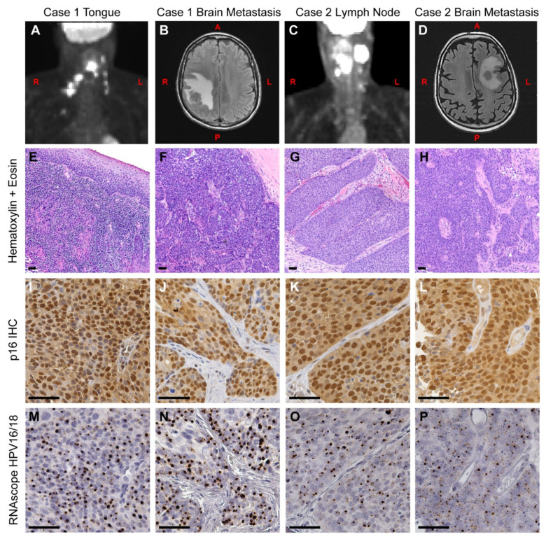
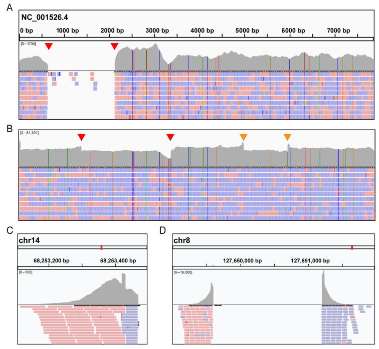
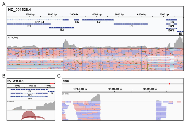
Human papillomavirus (HPV) positive and negative head and neck squamous cell carcinoma (HNSCC) are known to have differential phenotypes, including the incidence and location of metastases. HPV positive (HPV+) HNSCC are more likely to metastasize to distant sites, such as the lung, brain, and skin. Among these locations, metastasis to the brain is a rare event, and little is known about specific risk factors for this phenotype. In this report, we describe two patients who developed brain metastases from HNSCC. Both patient tumors had p16INK4a overexpression, suggesting these tumors were HPV+. This was confirmed after PCR, in situ hybridization, and mass spectrometry detected the presence of HPV type 16 (HPV16) DNA, RNA and protein. To further characterize the presence of HPV16, we used a target enrichment strategy on tumor DNA and RNA to isolate the viral sequences from the brain metastases. Analysis by targeted next generation sequencing revealed that both tumors had the HPV genome integrated into the host genome at known hotspots, 8q24.21 and 14q24.1. Applying a similar target enrichment strategy to a larger cohort of HPV+ HNSCC brain metastases could help to identify biomarkers that can predict metastasis and/or identify novel therapeutic options.

Keywords: DNA target enrichment; HNSCC; HPV; OPSCC; brain metastasis; data-independent acquisition; proteomics; targeted sequencing.


Conflict of interest statement

B.C.S. is a founder and shareholder in Proteome Software, which operates in the field of proteomics. The other authors declare no conflict of interest. The funders had no role in the design of the study; in the collection, analyses, or interpretation of data; in the writing of the manuscript, or in the decision to publish the results.

Figures

Figure 1
Figure 1
Clinical presentation and histology of primary and metastatic OPSCC. (A) A positron-emission tomography (PET) scan of Case 1 at the time of initial diagnosis, showing the primary tumor and spread to local lymph nodes. (B) A magnetic resonance imaging (MRI) scan was taken 16 months later, showing a right parietal mass in the brain. (C) A PET scan of Case 2 at the time of initial diagnosis, showing the neck mass and local spread to lymph nodes. (D) An MRI was taken 33 months later, showing a metastatic tumor in the left centrum. (EP) Serial tissue sections from Case 1 (E,F,I,J,M,N) and Case 2 (G,H,K,L,O,P) were used for immunohistochemistry (IHC) and in situ hybridization analysis. (EH) Primary and metastatic brain tumors were stained with hematoxylin and eosin. (IL) Adjacent sections were assayed for p16INK4a protein expression by IHC. All tumors displayed high levels of nuclear and cytoplasmic staining of p16INK4a, a hallmark of HPV+ tumors. (MP) Tumors were assayed for HPV16 and HPV18 RNA using the RNAscope HPV16/18 probe set. The presence of HPV RNA is evident in both initial (M,O) and metastatic brain tumors (N,P). This pattern directly matches the pattern of p16INK4a overexpression from the adjacent slides. L = left side, R = right side, A = anterior, P = posterior. Scale bar = 50 μm.
Figure 2
Figure 2
Characterization of human papillomavirus (HPV) integration in brain metastases by targeted sequencing. After sequencing the targeted libraries, reads were aligned to HPV16 reference genome (NC_001526.4) and visualized by IGV software. (A,B) Extensive coverage of the HPV genome was seen in both Case 1 (A) and Case 2 (B). Red arrows indicate discontinuities where chimeric reads between HPV and human genomes were detected. In addition, other discontinuities were detected in Case 2 (orange arrows). (C,D) In the lower panels, human DNA reads from chimeric HPV-human sequences are displayed, indicating an insertion site into the human genome at 14q24.1 (Case 1, (C)) and 8q24.21 (Case 2, (D)). Red (Forward) and blue (Reverse) colors correspond to specific strands.
Figure 3
Figure 3
Detection of HPV mRNA in Case 2 by targeted sequencing. Total RNA was first extracted from the Case 2 brain metastasis, followed by separation of the mRNA fraction using oligo dT magnetic beads. This was followed by cDNA library preparation and enrichment of HPV encoded cDNAs using the SureSelect protocols. After Illumina sequencing, reads were aligned to HPV16 reference genome (NC_001526.4) and visualized by IGV software. (A) Data revealed a high level of E6 and E7 expression in the tumor. (B) The junction track showing alternative splice junctions in the E6 gene. The E6*I splice junction had more coverage (8368 reads) than E6*II (1561 reads). (C) Consistent with the DNA sequencing, human reads were detected for the integration site at 8q24.21, indicating the presence of a chimeric mRNA encoded from both HPV16 and the host genome.

References

    1. Siegel R.L., Miller K.D., Jemal A. Cancer statistics, 2020. CA Cancer J. Clin. 2020;70:7–30. doi: 10.3322/caac.21590. - DOI - PubMed
    1. Vidal L., Gillison M.L. Human papillomavirus in HNSCC: Recognition of a distinct disease type. Hematol. Oncol. Clin. N. Am. 2008;22:1125–1142. doi: 10.1016/j.hoc.2008.08.006. vii. - DOI - PubMed
    1. Ang K.K., Harris J., Wheeler R., Weber R., Rosenthal D.I., Nguyen-Tan P.F., Westra W.H., Chung C.H., Jordan R.C., Lu C., et al. Human papillomavirus and survival of patients with oropharyngeal cancer. N. Engl. J. Med. 2010;363:24–35. doi: 10.1056/NEJMoa0912217. - DOI - PMC - PubMed
    1. Fakhry C., Westra W.H., Li S., Cmelak A., Ridge J.A., Pinto H., Forastiere A., Gillison M.L. Improved survival of patients with human papillomavirus-positive head and neck squamous cell carcinoma in a prospective clinical trial. J. Natl. Cancer Inst. 2008;100:261–269. doi: 10.1093/jnci/djn011. - DOI - PubMed
    1. Gillison M.L., D’Souza G., Westra W., Sugar E., Xiao W., Begum S., Viscidi R. Distinct risk factor profiles for human papillomavirus type 16-positive and human papillomavirus type 16-negative head and neck cancers. J. Natl. Cancer Inst. 2008;100:407–420. doi: 10.1093/jnci/djn025. - DOI - PubMed

Publication types

MeSH terms