Review
. 2021 Aug 6;13(8):1561.
doi: 10.3390/v13081561.

Hantavirus Replication Cycle-An Updated Structural Virology Perspective

Affiliations
Review

Hantavirus Replication Cycle-An Updated Structural Virology Perspective

Kristina Meier et al. Viruses. .

Erratum in

Abstract

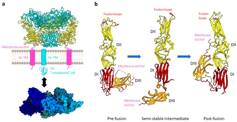
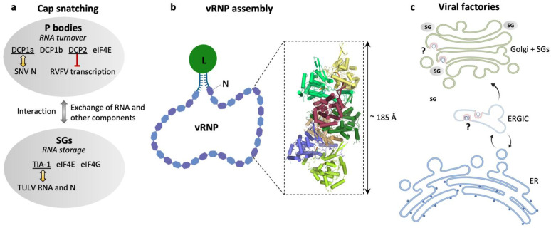
Hantaviruses infect a wide range of hosts including insectivores and rodents and can also cause zoonotic infections in humans, which can lead to severe disease with possible fatal outcomes. Hantavirus outbreaks are usually linked to the population dynamics of the host animals and their habitats being in close proximity to humans, which is becoming increasingly important in a globalized world. Currently there is neither an approved vaccine nor a specific and effective antiviral treatment available for use in humans. Hantaviruses belong to the order Bunyavirales with a tri-segmented negative-sense RNA genome. They encode only five viral proteins and replicate and transcribe their genome in the cytoplasm of infected cells. However, many details of the viral amplification cycle are still unknown. In recent years, structural biology methods such as cryo-electron tomography, cryo-electron microscopy, and crystallography have contributed essentially to our understanding of virus entry by membrane fusion as well as genome encapsidation by the nucleoprotein. In this review, we provide an update on the hantavirus replication cycle with a special focus on structural virology aspects.

Keywords: X-ray crystallography; cryo-electron microscopy; cryo-electron tomography; hantaviruses; structural virology; viral fusion glycoproteins; viral genome encapsidation; viral replication; viral transcription; virion assembly.


Conflict of interest statement

The authors declare no conflict of interest.

Figures

Figure 1
Figure 1
The hantavirus virion. A drawing of a hantavirus virion is presented (left) including a top view of the map of the TULV glycoprotein lattice (right), which was obtained by electron cryo-tomography and subvolume averaging (EMD-11236). A model of the prefusion tetrameric Gn/Gc spike complex (PDB: 6ZJM), presented as a cartoon, was fitted into the central volume [24].
Figure 2
Figure 2
The hantavirus spike complex. (a) A pre-fusion spike complex on the virus surface composed of the Gn (teal) and Gc (yellow) proteins forming a tetrameric assembly (example of ANDV, PDB: 6ZJM) is presented as a cartoon. The viral envelope (grey membrane) as well as the transmembrane regions (TM) of Gn (cyan cylinder) and Gc (pink cylinder) are schematically shown. One copy of the Gn cytoplasmic tail (cyan, PDB: 2K9H) and a heptameric RNP-like assembly of the N protein (blue; PDB: 6I2N) are displayed as a cartoon and surface representation, respectively. An interaction between the latter is indicated (black arrow). (b) A comparison of pre-fusion ANDV Gc (PDB: 6Y5F), an intermediate state of HTNV stabilized by an antibody (antibody not shown) (PDB: 5LJY), and the ANDV Gc post-fusion conformation (PDB: 6Y6Q) is shown. Domains are colored as DI, red; DII, yellow; DIII, orange. Key residues W766, Y745, F900 for ANDV and F250 for HTNV of the tripartite fusion loop are highlighted in red with the side chains displayed as sticks.
Figure 3
Figure 3
Hantavirus ribonucleoproteins and the location of viral genome replication and transcription. (a) Schematic representation of cellular RNA granules that have been reported as sites for hantavirus cap snatching: processing (P) bodies [86] and stress granules (SGs) [89]. Selected marker proteins for both granules are shown as well as eIF4E to emphasize its presence in both granules. Yellow arrows indicate reported colocalization of P body marker DCP1a and SNV N [86] or SGs marker TIA-1 and TULV RNA and N [89], and the red barred arrow indicates inhibition of RVFV transcription by P body-resident protein DCP2 [88]. (b) Schematic representation of a viral ribonucleoprotein (vRNP). The viral RNA is associated with N and L proteins in a panhandle-like conformation with the complementary genome ends forming a partially double-stranded region and the termini is most probably bound to the L protein. The close-up represents a heptameric assembly of N (PDB: 6I2N) into a helical RNP-like structure with tri-nucleotide RNAs (orange) bound to each N protomer. (c) Overview of possible locations of viral factories for genome transcription and replication in the endoplasmic reticulum (ER), endoplasmic reticulum–Golgi intermediate compartment (ERGIC) [100], and the Golgi apparatus associated with SGs [89].

References

    1. Maes P., Alkhovsky S.V., Bao Y., Beer M., Birkhead M., Briese T., Buchmeier M.J., Calisher C.H., Charrel R.N., Choi I.R., et al. Taxonomy of the family Arenaviridae and the order Bunyavirales: Update 2018. Arch. Virol. 2018;163:2295–2310. doi: 10.1007/s00705-018-3843-5. - DOI - PubMed
    1. Avsic-Zupanc T., Saksida A., Korva M. Hantavirus infections. Clin. Microbiol. Infect. 2019;21S:e6–e16. doi: 10.1111/1469-0691.12291. - DOI - PubMed
    1. Vaheri A., Henttonen H., Voutilainen L., Mustonen J., Sironen T., Vapalahti O. Hantavirus infections in Europe and their impact on public health. Rev. Med. Virol. 2013;23:35–49. doi: 10.1002/rmv.1722. - DOI - PubMed
    1. Hjelle B., Torres-Perez F. Hantaviruses in the americas and their role as emerging pathogens. Viruses. 2010;2:2559–2586. doi: 10.3390/v2122559. - DOI - PMC - PubMed
    1. Padula P.J., Edelstein A., Miguel S.D.L., Lopez N.M., Rossi C.M., Rabinovich R.D. Hantavirus Pulmonary Syndrome Outbreak in Argentina: Molecular Evidencefor Person-to-Person Transmission of Andes Virus. Virology. 1998 doi: 10.1006/viro.1997.8976. - DOI - PubMed

Publication types

LinkOut - more resources