

Acute Late-Stage Myocarditis in the Crab-Eating Macaque Model of Hemorrhagic Smallpox
- PMID: 34452435
- PMCID: PMC8402688
- DOI: 10.3390/v13081571
Acute Late-Stage Myocarditis in the Crab-Eating Macaque Model of Hemorrhagic Smallpox
Abstract
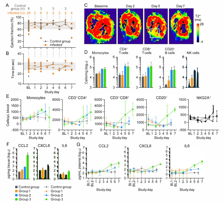
Hemorrhagic smallpox, caused by variola virus (VARV), was a rare but nearly 100% lethal human disease manifestation. Hemorrhagic smallpox is frequently characterized by secondary bacterial infection, coagulopathy, and myocardial and subendocardial hemorrhages. Previous experiments have demonstrated that intravenous (IV) cowpox virus (CPXV) exposure of macaques mimics human hemorrhagic smallpox. The goal of this experiment was to further understand the onset, nature, and severity of cardiac pathology and how it may contribute to disease. The findings support an acute late-stage myocarditis with lymphohistiocytic infiltrates in the CPXV model of hemorrhagic smallpox.
Keywords: CPXV; VARV; biodefense; cardiac MRI; cowpox; myocarditis; smallpox; variola.
Conflict of interest statement
The authors declare no conflict of interest. The funders had no role in the design of the study; in the collection, analyses, or interpretation of data; in the writing of the manuscript, or in the decision to publish the results. The content of this publication does not necessarily reflect the views or policies of the U.S. Department of Health and Human Services (HHS) or of the institutions and companies affiliated with the authors. Mention of trade names, commercial products, or organizations does not imply endorsement by the U.S. Government.
Figures
Similar articles
-
Cowpox virus infection of cynomolgus macaques as a model of hemorrhagic smallpox.Virology. 2011 Sep 30;418(2):102-12. doi: 10.1016/j.virol.2011.07.013. Epub 2011 Aug 15. Virology. 2011. PMID: 21840027 Free PMC article.
-
Intrabronchial inoculation of cynomolgus macaques with cowpox virus.J Gen Virol. 2012 Jan;93(Pt 1):159-164. doi: 10.1099/vir.0.036905-0. Epub 2011 Sep 21. J Gen Virol. 2012. PMID: 21940414 Free PMC article.
-
Progression of pathogenic events in cynomolgus macaques infected with variola virus.PLoS One. 2011;6(10):e24832. doi: 10.1371/journal.pone.0024832. Epub 2011 Oct 6. PLoS One. 2011. PMID: 21998632 Free PMC article.
-
Modulation of the host immune response by cowpox virus.Microbes Infect. 2010 Nov;12(12-13):900-9. doi: 10.1016/j.micinf.2010.07.007. Epub 2010 Jul 29. Microbes Infect. 2010. PMID: 20673807 Free PMC article. Review.
-
[Clinical presentation of cowpox virus infection in South American camelids - A review].Tierarztl Prax Ausg G Grosstiere Nutztiere. 2018 Feb;46(1):50-56. doi: 10.15653/TPG-170502. Epub 2018 Feb 21. Tierarztl Prax Ausg G Grosstiere Nutztiere. 2018. PMID: 29536472 Review. German.
Cited by
-
Myocarditis Attributable to Monkeypox Virus Infection in 2 Patients, United States, 2022.Emerg Infect Dis. 2022 Dec;28(12):2508-2512. doi: 10.3201/eid2812.221276. Epub 2022 Sep 30. Emerg Infect Dis. 2022. PMID: 36179413 Free PMC article.
-
Cardiac Involvement in Monkeypox Outbreak.Discoveries (Craiova). 2023 Sep 18;11(3):e171. doi: 10.15190/d.2023.10. eCollection 2023 Jul-Sep. Discoveries (Craiova). 2023. PMID: 37753488 Free PMC article. Review.
References
-
- Henderson D.A. Smallpox: The Death of a Disease. The Inside Story of Eradicating a Worldwide Killer. Prometheus; Amherst, NY, USA: 2009.
-
- US Department of Health and Human Services. Public Health Service. Centers for Disease Control and Prevention. National Institutes of Health Meechan P.J., Potts J., editors. [(accessed on 1 December 2020)];Biosafety in Microbiological and Biomedical Laboratories. (6th ed.). 2020 Available online: https://www.cdc.gov/labs/BMBL.html.
-
- Rao A.R. Clinical Smallpox. Classification and Frequency of Type of Variola Major Document SE/72.1. World Health Organization; Geneva, Switzerland: 1972.
-
- Rao A.R. Haemorrhagic smallpox: A study of 240 cases. J. Indian Med. Assoc. 1964;43:224–229. - PubMed
Publication types
MeSH terms
Grants and funding
LinkOut - more resources
Full Text Sources
Medical
