. 2021 Aug 11;13(8):1587.
doi: 10.3390/v13081587.

Interaction of Poliovirus Capsid Proteins with the Cellular Autophagy Pathway

Affiliations

Interaction of Poliovirus Capsid Proteins with the Cellular Autophagy Pathway

Anna Zimina et al. Viruses. .

Abstract

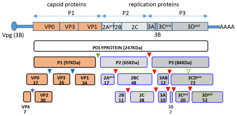
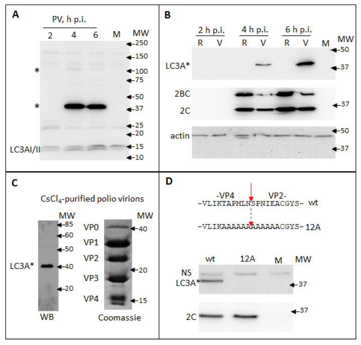
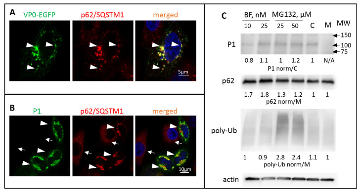
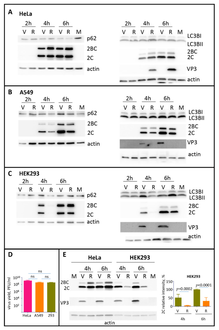
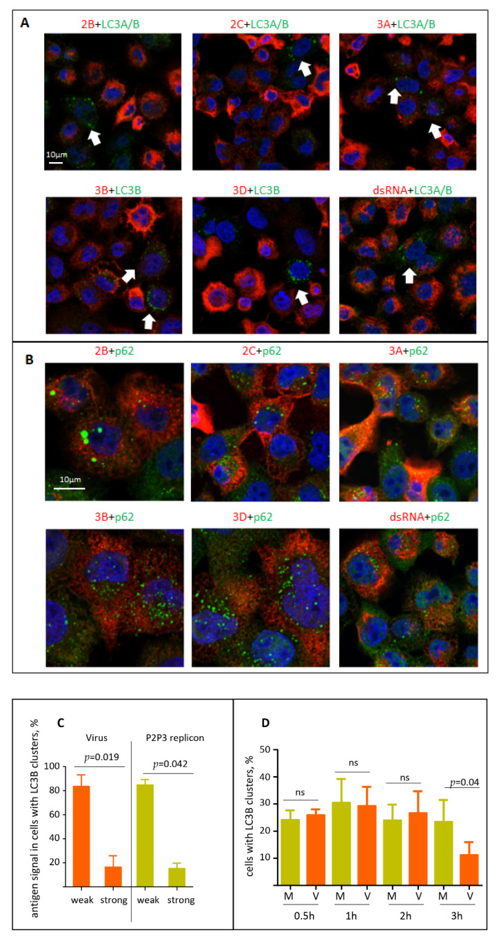
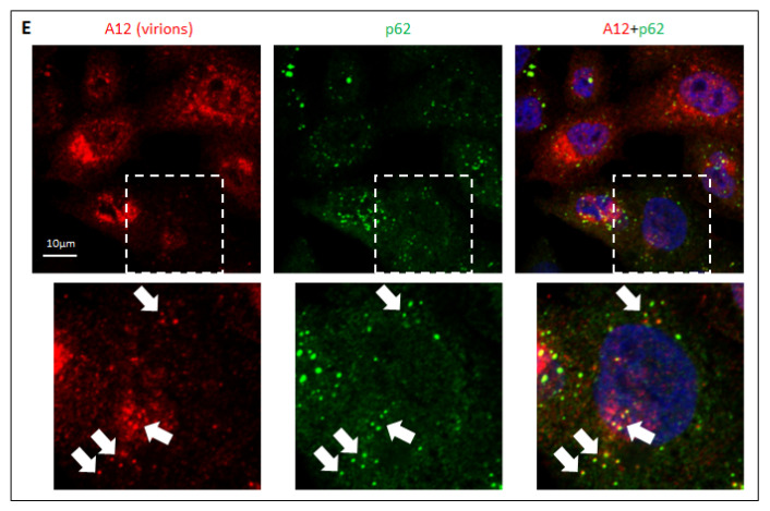
The capsid precursor P1 constitutes the N-terminal part of the enterovirus polyprotein. It is processed into VP0, VP3, and VP1 by the viral proteases, and VP0 is cleaved autocatalytically into VP4 and VP2. We observed that poliovirus VP0 is recognized by an antibody against a cellular autophagy protein, LC3A. The LC3A-like epitope overlapped the VP4/VP2 cleavage site. Individually expressed VP0-EGFP and P1 strongly colocalized with a marker of selective autophagy, p62/SQSTM1. To assess the role of capsid proteins in autophagy development we infected different cells with poliovirus or encapsidated polio replicon coding for only the replication proteins. We analyzed the processing of LC3B and p62/SQSTM1, markers of the initiation and completion of the autophagy pathway and investigated the association of the viral antigens with these autophagy proteins in infected cells. We observed cell-type-specific development of autophagy upon infection and found that only the virion signal strongly colocalized with p62/SQSTM1 early in infection. Collectively, our data suggest that activation of autophagy is not required for replication, and that capsid proteins contain determinants targeting them to p62/SQSTM1-dependent sequestration. Such a strategy may control the level of capsid proteins so that viral RNAs are not removed from the replication/translation pool prematurely.

Keywords: LC3 processing; autophagy; enterovirus replication; enteroviruses; poliovirus.


Conflict of interest statement

The authors declare no conflict of interest.

Figures

Figure 1
Figure 1
Scheme of poliovirus genome organization and polyprotein processing. Cleavage sites of the protease 2A are marked by green, those of 3C by red, and those of 3CD by blue triangles, respectively. Purple star denotes the autocatalytic cleavage site between VP4 and VP2. Numbers indicate the molecular weight of the corresponding proteins in KDa.
Figure 2
Figure 2
Poliovirus capsid protein VP0 contains an LC3A-like epitope. (A) HeLa cells were infected with poliovirus at an MOI of 10 and the samples were collected at the indicated times post-infection. Mock-infected sample is collected at 6 h. Western blot was developed with a rabbit monoclonal anti-LC3A antibody (Cell Signaling). LC3A * designates abnormal higher molecular weight LC3A signals. (B) HeLa cells were transfected with either full-length poliovirus RNA (V) or replicon RNA (R) coding for only P2P3 replication proteins and developed with the same anti-LC3A antibody as in A (upper panel). Staining with anti-polio 2C antibody shows polio replication (middle panel). Actin is shown as a loading control (lower panel). (C) Proteins from poliovirus virions purified through CsCl4 gradient were resolved on an SDS-PAGE gel and either subjected to a Western blotting with the anti-LC3A antibody (left), or visualized with a Coomassie stain (right). (D) Top—amino-acid sequences of the VP4/VP2 cleavage site in VP0 and the alanine substitutions in the 12A mutant. HeLa cells were transfected with either wt polio RNA or the RNA with 12A substitution and the cell lysates were subject to Western blots with either anti-LC3A (top panel) or anti-polio 2C antibodies (bottom panel).
Figure 3
Figure 3
Poliovirus capsid proteins are sequestered in a p62/SQSTM-dependent manner. (A) HeLa cells expressing VP0-EGFP fusion (green) stained with an anti- p62/SQSTM antibody (red) Arrowheads show colocalization of both signals. Nuclear DNA is stained with Hoechst 33,342 (blue). (B) HeLa cells expressing the whole capsid protein precursor P1 stained with anti-polio VP1 (green) and anti- p62/SQSTM (red) antibodies. Arrowheads and arrows show cells that either express or do not express P1, respectively. Note the concentration of p62/SQSTM signal in the former compared to the mostly diffuse staining in the latter. (C) HeLa cells were transfected with a plasmid expressing the whole P1 capsid protein precursor, and at 18 h post-transfection an inhibitor of lysosome acidification bafilomycin or a proteasome inhibitor MG132 were added at the indicated concentrations. C-control cells not treated with inhibitors. Total cell lysate was prepared after six hours of incubation with the inhibitors and analyzed with the anti-VP3 antibodies in a Western blot to assess the accumulation of P1 and with anti-p62 and anti-ubiquitin antibodies to confirm the efficacy of the inhibitor treatment. Quantitation shows P1 signal normalized to that in the sample not treated with inhibitors (C), p62 and polyubiquitin signals are normalized to those in the sample transfected with an empty vector (M). Actin is shown as a loading control.
Figure 4
Figure 4
The development of autophagy upon polio infection is cell type specific. (AC) HeLa, A549, or HEK293 cells, respectively, were infected with the MOI of 25 of either poliovirus (V) or encapsidated P2P3 replicon (R), and the cells were lysed at the indicated times post-infection. Lysates from mock-infected cells were prepared at 6 h. The same lysates were resolved either on 12% (left panels) or on 4–15% gradient gels (right panels) and analyzed in Western blots with the indicated antibodies. Staining with anti-2C antibody shows polio replication, staining with anti-VP3 antibody confirms the absence of capsid protein expression in cells infected with the replicon construct, actin is shown as a loading control. (D) HeLa, A549 and HEK 293 cells were infected with 25PFU of poliovirus and the virus yield at 6 h p.i. was determined by plaque assays. The difference in the titers between any of the samples was non-significant (ns). (E) HEK293 and HeLa cells were infected in parallel with the same preparations of either poliovirus (V) or encapsidated P2P3 replicon (R), collected at the indicated times post-infection and analyzed in a Western blot with anti-polio 2C and VP3 (capsid protein) antibodies. Actin is shown as a loading control. Note a significantly compromised replication of replicon compared to poliovirus in HEK293 but not HeLa cells. Quantitation shows 2C signals normalized to that in the virus-infected sample at 6 h p.i. from three independent experiments of infection of HEK293 cells with encapsidated replicon and poliovirus.
Figure 5
Figure 5
Localization of the viral antigens and autophagy proteins in infected cells. (A) HeLa cells were infected with an MOI of 25 of encapsidated P2P3 replicon, fixed at 4 h p.i. and processed for immunofluorescence with either rabbit monoclonal anti-LC3A/B or mouse monoclonal anti-LC3B (green) antibodies and the indicated polio antigens (red). Nuclear DNA is stained with Hoechst 33,342 (blue). (B) Same as in A but with either rabbit polyclonal or mouse monoclonal anti- p62/SQSTM (green) antibodies. (C) HeLa cells were infected with an MOI of 25 of either encapsidated P2P3 replicon or poliovirus, and at 4 h p.i. stained with antibodies against polio antigens 3D or 3B (rabbit polyclonal), and LC3B (mouse monoclonal). The level of viral antigen expression was quantified in at least 50 cells with LC3B clusters from multiple random fields of view. (D) HeLa cells were infected (V) or mock-infected (M) with an MOI of 25 of poliovirus, and at the indicated time post infection stained for LC3B. The percentage of cells with LC3B clusters was quantified from at least 100 cells per sample from multiple random fields of view. (E) HeLa cells were infected with the MOI of 25 of poliovirus, fixed at 4 h p.i. and processed for immunofluorescence with a monoclonal antibody A12 (red) recognizing assembled polio virions or virus-like particles and an antibody against p62/SQSTM (green). Nuclear DNA is stained with Hoechst 33,342 (blue). Arrows show colocalization of A12 and p62/SQSTM signals in cells at the early stages of virion accumulation (the selected area is shown).
Figure 5
Figure 5
Localization of the viral antigens and autophagy proteins in infected cells. (A) HeLa cells were infected with an MOI of 25 of encapsidated P2P3 replicon, fixed at 4 h p.i. and processed for immunofluorescence with either rabbit monoclonal anti-LC3A/B or mouse monoclonal anti-LC3B (green) antibodies and the indicated polio antigens (red). Nuclear DNA is stained with Hoechst 33,342 (blue). (B) Same as in A but with either rabbit polyclonal or mouse monoclonal anti- p62/SQSTM (green) antibodies. (C) HeLa cells were infected with an MOI of 25 of either encapsidated P2P3 replicon or poliovirus, and at 4 h p.i. stained with antibodies against polio antigens 3D or 3B (rabbit polyclonal), and LC3B (mouse monoclonal). The level of viral antigen expression was quantified in at least 50 cells with LC3B clusters from multiple random fields of view. (D) HeLa cells were infected (V) or mock-infected (M) with an MOI of 25 of poliovirus, and at the indicated time post infection stained for LC3B. The percentage of cells with LC3B clusters was quantified from at least 100 cells per sample from multiple random fields of view. (E) HeLa cells were infected with the MOI of 25 of poliovirus, fixed at 4 h p.i. and processed for immunofluorescence with a monoclonal antibody A12 (red) recognizing assembled polio virions or virus-like particles and an antibody against p62/SQSTM (green). Nuclear DNA is stained with Hoechst 33,342 (blue). Arrows show colocalization of A12 and p62/SQSTM signals in cells at the early stages of virion accumulation (the selected area is shown).

Similar articles

Cited by

References

    1. Ypmawong M.F., Dewalt P.G., Johnson V.H., Lamb J.G., Semler B.L. Protein 3cd Is the Major Poliovirus Proteinase Responsible for Cleavage of the P1 Capsid Precursor. Virology. 1988;166:265–270. doi: 10.1016/0042-6822(88)90172-9. - DOI - PubMed
    1. Toyoda H., Nicklin M.J., Murray M.G., Anderson C.W., Dunn J.J., Studier F.W., Wimmer E. A second virus-encoded proteinase involved in proteolytic processing of poliovirus polyprotein. Cell. 1986;45:761–770. doi: 10.1016/0092-8674(86)90790-7. - DOI - PubMed
    1. Putnak J.R., Phillips B.A. Picornaviral structure and assembly. Microbiol. Rev. 1981;45:287–315. doi: 10.1128/mr.45.2.287-315.1981. - DOI - PMC - PubMed
    1. Fields B.N., Knipe D.M., Howley P.M. Fields Virology. 5th ed. Wolters Kluwer Health/Lippincott Williams & Wilkins; Philadelphia, PA, USA: 2007.
    1. Leveque N., Semler B.L. A 21st Century Perspective of Poliovirus Replication. PLoS Pathog. 2015;11 doi: 10.1371/journal.ppat.1004825. - DOI - PMC - PubMed

Publication types

LinkOut - more resources