. 2021 Aug 11;13(8):1589.
doi: 10.3390/v13081589.

Efficient Transendothelial Migration of Latently HIV-1-Infected Cells

Affiliations

Efficient Transendothelial Migration of Latently HIV-1-Infected Cells

Reou Tanabe et al. Viruses. .

Abstract

A small fraction of HIV-1-infected T cells forms populations of latently infected cells when they are a naive T-cell subset or in transit to a resting memory state. Latently HIV-1-infected cells reside in lymphoid tissues and serve as viral reservoirs. However, whether they systemically recirculate in the body and re-enter the lymphoid nodes are unknown. Here, we employed two in-vitro cell coculture systems mimicking the lymphatic endothelium in lymph nodes and investigated the homing potential, specifically the transendothelial migration (TEM), of two latently HIV-1-infected cell lines (J1.1 and ACH-2). In trans-well coculture systems, J1.1 and ACH-2 showed higher TEM efficiencies than their parental uninfected and acutely infected cells. The efficiency of TEM was enhanced by the presence of stromal cells, such as HS-5 and fibroblastic reticular cells. In an in-vitro reconstituted, three-dimensional coculture system in which stromal cells are embedded in collagen matrices, J1.1 showed slightly higher TEM efficiency in the presence of HS-5. In accordance with these phenotypes, latently infected cells adhered to the endothelial cells more efficiently than uninfected cells. Together, our study showed that latently HIV-1-infected cells enhanced cell adhesion and TEM abilities, suggesting their potential for efficient homing to lymph nodes.

Keywords: HIV-1; latent infection; lymphocyte homing; reconstitution; stromal cells; transmigration.


Conflict of interest statement

The authors declare no conflict of interest.

Figures
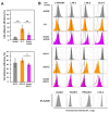
Figure 1
Figure 1
Cell adhesion ability and adhesion molecule expression of latently and acutely infected T cells. (A) Adhesion ability of Jurkat and A3.01 cell lineages. Uninfected, latently infected, and acutely infected cells of Jurkat (upper) and A3.01 (lower) cell lineages were labeled with CellTracker CMFDA. They were added to EA.hy926 cell monolayers and incubated for 1 h at 37 °C. After washing, the cells were lysed with RIPA buffer (1 mL). The MFI of the lysates (200 μL) was measured at 480 nm/520 nm and corrected using background control samples. Data are the mean ± S.D. (n = 15) from three independent experiments. * p < 0.05; ** p < 0.01. (B) Expression of cell adhesion molecules of Jurkat and A3.01 cell lineages. Uninfected cells, latently infected cells, and acutely infected cells of Jurkat and A3.01 cell lineages were immune-stained with anti-L-selection, anti-LFA-1, anti-LFA-2, and anti-VLA-4 mAbs and analyzed by FCM. EA.hy926 cells were immune-stained with anti-ICAM-1, anti-VCAM-1, anti-PECAM-1, and anti-LFA-3 mAb and subjected to FCM. White histograms show negative controls with secondary Ab alone. Dashed lines indicate peaks of their expressions in parental Jurkat and A3.01 cells.
Figure 2
Figure 2
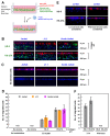
TEM efficiency of latently infected and acutely infected T cells in transwell systems. (A) A TEM system in transwell plates. EA.hy926 cells were grown in cell inserts of transwell plates (upper chambers), whereas stromal HS-5 or FRC were separately grown in plate wells (lower chambers). They were combined just before use. Uninfected, latently infected, and acutely infected T cells were labeled with CellTracker CMFDA. They were added to the EA.hy926 cell monolayers and incubated for 6 h at 37 °C in the presence of chemokines (CXCL12, CCL19, and CCL21). Transmigrated cells were collected and lysed with RIPA buffer (1 mL). The MFI of the lysates (200 μL) was measured at 480 nm/520 nm and corrected using background control samples. (B) TEM of Jurkat (upper) and A3.01 (lower) cell lineages across EA.hy926 cell monolayers. CMFDA-labeled T cells were added to the EA.hy926 cell monolayers in cell inserts, but stromal cells were absent in plate wells. Data are the mean ± S.D. from 4 or 5 independent experiments (upper) and 5 or 6 independent experiments (lower). * p < 0.05; ** p < 0.01. (C) TEM of Jurkat cell lineage (uninfected, latently infected, and acutely infected) through EA.hy926 cell monolayers in the absence (-) or presence of stromal cells (HS-5 and FRC) in plate wells. Data are the mean ± S.D. from at least three independent experiments. * p < 0.05.
Figure 3
Figure 3
TEM of latently infected and acutely infected T cells in in-vitro reconstructed lymphatic endothelium systems. (A) In-vitro reconstituted lymphatic endothelium systems with collagen. Stromal cells were labeled with CellTrace CFSE and were embedded in reconstituted collagen gels with chemokines (CCL19, CCL21 and CXCL12). EA.hy926 cells were labeled with CellTracker CMTMR and grown to monolayers on the collagen gels. Uninfected Jurkat, J1.1, and acutely infected Jurkat cells were labeled with Hoechst 33342, added to the EA.hy926 monolayers, and incubated at 37 °C for 3 h. The z-stack images were collected and were processed to generate 3-D reconstituted images using Fiji in ImageJ software. (B,C) TEM of Jurkat cell lineage into the collagen gels. HS-5 and HS-27A were embedded (B) or not embedded (C) in the reconstituted collagen gels. EA.hy926 cells were present in ((C), upper) but absent in ((C), lower). (E) TEM of Jurkat cells into the collagen gels embedded with live (left) or PFA-fixed (right) stromal cells. The stromal cells were not labeled with CellTrace CFSE. (D,F) For quantification, the number of migrated cells was calculated in a z-projection image. Data are the mean ± S.D. from three independent experiments. N.D., not detectable.

References

    1. Finzi D., Hermankova M., Pierson T., Carruth L.M., Buck C., Chaisson R.E., Quinn T.C., Chadwick K., Margolick J., Brookmeyer R., et al. Identification of a reservoir for HIV-1 in patients on highly active antiretroviral therapy. Science. 1997;278:1295–1300. doi: 10.1126/science.278.5341.1295. - DOI - PubMed
    1. Wong J.K., Hezareh M., Günthard H.F., Havlir D.V., Ignacio C.C., Spina C.A., Richman D.D. Recovery of replication-competent HIV despite prolonged suppression of plasma viremia. Science. 1997;278:1291–1295. doi: 10.1126/science.278.5341.1291. - DOI - PubMed
    1. Stevenson M. HIV-1 pathogenesis. Nat. Med. 2003;9:853–861. doi: 10.1038/nm0703-853. - DOI - PubMed
    1. Sengupta S., Siliciano R.F. Targeting the Latent Reservoir for HIV-1. Immunity. 2018;48:872–895. doi: 10.1016/j.immuni.2018.04.030. - DOI - PMC - PubMed
    1. Wong J.K., Yukl S.A. Tissue reservoirs of HIV. Curr. Opin. HIV AIDS. 2016;11:362–370. doi: 10.1097/COH.0000000000000293. - DOI - PMC - PubMed

Publication types

Substances

LinkOut - more resources