. 2021 Aug 11;13(8):1592.
doi: 10.3390/v13081592.

Genomic Epidemiology and Evolution of Duck Hepatitis A Virus

Affiliations

Genomic Epidemiology and Evolution of Duck Hepatitis A Virus

Enikő Fehér et al. Viruses. .

Abstract

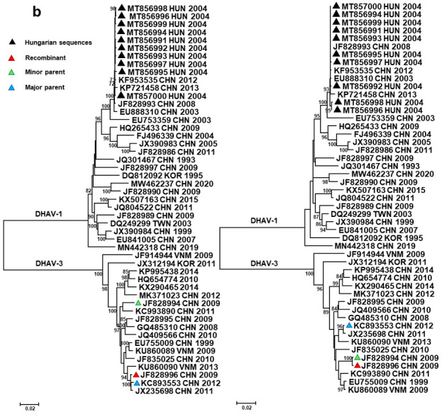
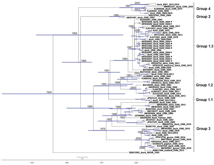
Duck hepatitis A virus (DHAV), an avian picornavirus, causes high-mortality acute disease in ducklings. Among the three serotypes, DHAV-1 is globally distributed, whereas DHAV-2 and DHAV-3 serotypes are chiefly restricted to Southeast Asia. In this study, we analyzed the genomic evolution of DHAV-1 strains using extant GenBank records and genomic sequences of 10 DHAV-1 strains originating from a large disease outbreak in 2004-2005, in Hungary. Recombination analysis revealed intragenotype recombination within DHAV-1 as well as intergenotype recombination events involving DHAV-1 and DHAV-3 strains. The intergenotype recombination occurred in the VP0 region. Diversifying selection seems to act at sites of certain genomic regions. Calculations estimated slightly lower rates of evolution of DHAV-1 (mean rates for individual protein coding regions, 5.6286 × 10⁻⁴ to 1.1147 × 10⁻³ substitutions per site per year) compared to other picornaviruses. The observed evolutionary mechanisms indicate that whole-genome-based analysis of DHAV strains is needed to better understand the emergence of novel strains and their geographical dispersal.

Keywords: Hungary; duck hepatitis A virus; recombination.


Conflict of interest statement

The authors declare no conflict of interest. The funders had no role in the design of the study; in the collection, analyses, or interpretation of data; in the writing of the manuscript, or in the decision to publish the results.

Figures

Figure 1
Figure 1
Geographic distribution of DHAV serotypes/genotypes. DHV type I is a historic assignment of DHAV introduced prior to the separation of serotypes.
Figure 2
Figure 2
Unrooted neighbor-joining phylogenetic trees representing intergenotypic (a) and intragenotypic (b) recombination events. The trees were generated with a p-distance model (1000 bootstrap replicates) using sequence alignments extracted from polyprotein sequences according to the predicted recombination points determined with RDP4 software. The trees show groupings of recombinant strains with the putative major (tree on the left) and minor (tree on the right) parents (or related strains with GenBank records in all cases when true parents are unknown).
Figure 2
Figure 2
Unrooted neighbor-joining phylogenetic trees representing intergenotypic (a) and intragenotypic (b) recombination events. The trees were generated with a p-distance model (1000 bootstrap replicates) using sequence alignments extracted from polyprotein sequences according to the predicted recombination points determined with RDP4 software. The trees show groupings of recombinant strains with the putative major (tree on the left) and minor (tree on the right) parents (or related strains with GenBank records in all cases when true parents are unknown).
Figure 3
Figure 3
Results of the selection constraint analysis. β+ values (y-axis) at individual sites (x-axis) were extracted and plotted from results of the MEME analysis (green circles). Sites under positive selection predicted with high significance values are labeled with red triangles and the yellow square.
Figure 4
Figure 4
Time-calibrated maximum clade credibility tree of 102 VP1 sequences generated by the BEAST package 1.10.4, HKY + G model. Genetic groups and subgroups are on the right.

Similar articles

Cited by

References

    1. Hisham I., Ellakany H.F., Selim A.A., Abdalla M.A.M., El-Abideen M.A.Z., Kilany W.H., Ali A., Elbestawy A.R. Comparative pathogenicity of duck hepatitis A virus-1 isolates in experimentally infected Pekin and Muscovy ducklings. Front. Vet. Sci. 2020;7:234. doi: 10.3389/fvets.2020.00234. - DOI - PMC - PubMed
    1. Levine P.P., Fabricant J. A hitherto-undescribed virus disease of ducks in North America. Cornell Vet. 1950;40:71–86.
    1. Liu R., Shi S., Huang Y., Chen Z., Chen C., Cheng L., Fu G., Chen H., Wan C., Fu Q. Comparative pathogenicity of different subtypes of duck hepatitis A virus in Pekin ducklings. Vet. Microbiol. 2019;228:181–187. doi: 10.1016/j.vetmic.2018.11.030. - DOI - PubMed
    1. Niu Y., Ma H., Ding Y., Li Z., Sun Y., Li M., Shi Y. The pathogenicity of duck hepatitis A virus types 1 and 3 on ducklings. Poult. Sci. 2019;98:6333–6339. doi: 10.3382/ps/pez455. - DOI - PMC - PubMed
    1. Niu Y., Liu B., Sun C., Zhao L., Chen H. Construction of the recombinant duck enteritis virus delivering capsid protein VP0 of the duck hepatitis A virus. Vet. Microbiol. 2020;249:108837. doi: 10.1016/j.vetmic.2020.108837. - DOI - PubMed

Publication types

LinkOut - more resources