

Scale Drop Disease Virus Associated Yellowfin Seabream (Acanthopagrus latus) Ascites Diseases, Zhuhai, Guangdong, Southern China: The First Description
- PMID: 34452481
- PMCID: PMC8402775
- DOI: 10.3390/v13081617
Scale Drop Disease Virus Associated Yellowfin Seabream (Acanthopagrus latus) Ascites Diseases, Zhuhai, Guangdong, Southern China: The First Description
Abstract
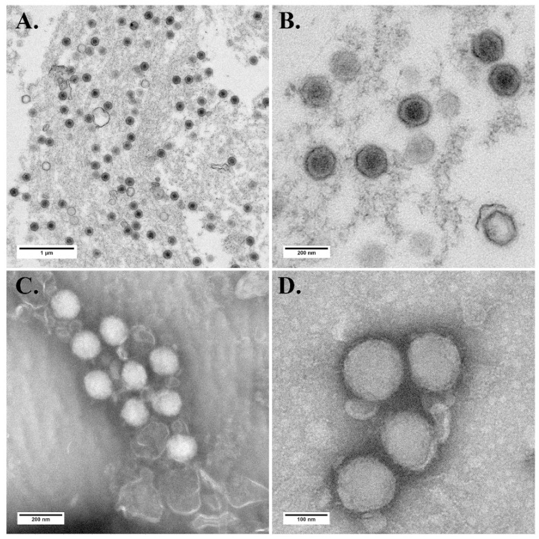
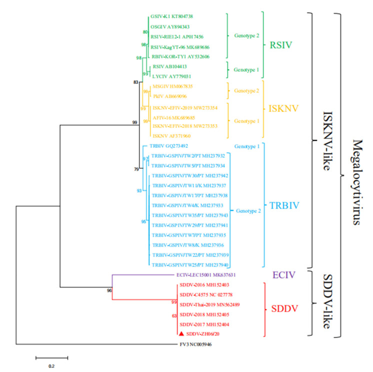
Scale drop disease virus (SDDV), an emerging piscine iridovirus prevalent in farmed Asian seabass Lates calcarifer in Southeast Asia, was firstly scientifically descripted in Singapore in 2015. Here, an SDDV isolate ZH-06/20 was isolated by inoculating filtered ascites from diseased juvenile yellowfin seabream into MFF-1 cell. Advanced cytopathic effects were observed 6 days post-inoculation. A transmission electron microscopy examination confirmed that numerous virion particles, about 140 nm in diameter, were observed in infected MFF-1 cell. ZH-06/20 was further purified and both whole genome and virion proteome were determined. The results showed that ZH-06/20 was composed of 131,122 bp with 135 putative viral proteins and 113 of them were further detected by virion proteome. Western blot analysis showed that no (or weak) cross-reaction was observed among several major viral proteins between ZH-06/20 and ISKNV-like megalocytivirus. An artificial challenge showed that ZH-06/20 could cause 100% death to juvenile yellowfin seabream. A typical sign was characterized by severe ascites, but not scale drop, which was considerably different from SDD syndrome in Asian seabass. Collectively, SDDV was confirmed, for the first time, as the causative agent of ascites diseases in farmed yellowfin seabream. Our study offers useful information to better understanding SDDV-associated diseases in farmed fish.
Keywords: genome; pathogenicity; proteome; scale drop disease virus; yellowfin seabream ascites diseases.
Conflict of interest statement
The authors declare no conflict of interest.
Figures


Similar articles
-
Detection and Genomic Characterisation of Scale Drop Disease Virus (SDDV) in Farmed Yellowfin Seabream (Acanthopagrus latus) in Southern China.J Fish Dis. 2025 Jun 20:e14161. doi: 10.1111/jfd.14161. Online ahead of print. J Fish Dis. 2025. PMID: 40539359
-
PCR Detection and Phylogenetic Analysis of Megalocytivirus Isolates in Farmed Giant Sea Perch Lates calcarifer in Southern Taiwan.Viruses. 2020 Jun 24;12(6):681. doi: 10.3390/v12060681. Viruses. 2020. PMID: 32599850 Free PMC article.
-
Mortality from scale drop disease in farmed Lates calcarifer in Southeast Asia.J Fish Dis. 2019 Jan;42(1):119-127. doi: 10.1111/jfd.12915. Epub 2018 Nov 5. J Fish Dis. 2019. PMID: 30397913
-
Megalocytiviruses.Viruses. 2012 Apr;4(4):521-38. doi: 10.3390/v4040521. Epub 2012 Apr 10. Viruses. 2012. PMID: 22590684 Free PMC article. Review.
-
[Red sea bream iridoviral disease].Uirusu. 2005 Jun;55(1):115-25. doi: 10.2222/jsv.55.115. Uirusu. 2005. PMID: 16308538 Review. Japanese.
Cited by
-
Comparative genomic analysis reveals new evidence of genus boundary for family Iridoviridae and explores qualified hallmark genes.Comput Struct Biotechnol J. 2022 Jun 27;20:3493-3502. doi: 10.1016/j.csbj.2022.06.049. eCollection 2022. Comput Struct Biotechnol J. 2022. PMID: 35860404 Free PMC article.
-
Fish Iridoviridae: infection, vaccination and immune response.Vet Res. 2024 Jul 15;55(1):88. doi: 10.1186/s13567-024-01347-1. Vet Res. 2024. PMID: 39010235 Free PMC article. Review.
-
Vitamin C Inhibits Scale Drop Disease Virus Infectivity by Targeting Nrf2 to Reduce Ferroptosis.Antioxidants (Basel). 2025 May 10;14(5):576. doi: 10.3390/antiox14050576. Antioxidants (Basel). 2025. PMID: 40427458 Free PMC article.
-
Development of a Double-Antibody Sandwich ELISA for Rapid Detection of the MCP Antigen Concentration in Inactivated ISKNV Vaccines.Vaccines (Basel). 2021 Nov 2;9(11):1264. doi: 10.3390/vaccines9111264. Vaccines (Basel). 2021. PMID: 34835196 Free PMC article.
-
Protective efficiency and immune responses to single and booster doses of formalin-inactivated scale drop disease virus (SDDV) vaccine in Asian seabass (Lates calcarifer).BMC Vet Res. 2024 Jun 20;20(1):267. doi: 10.1186/s12917-024-04132-6. BMC Vet Res. 2024. PMID: 38902724 Free PMC article.
References
-
- Girisha S.K., Kushala K.B., Nithin M.S., Puneeth T.G., Kumar B.T.N., Vinay T.N., Suresh T., Ajay S.K., Venugopal M.N., Ramesh K.S. First report of the infectious spleen and kidney necrosis virus (ISKNV) infection in ornamental fishes in India. Transbound. Emerg. Dis. 2021;68:964–972. doi: 10.1111/tbed.13793. - DOI - PubMed
Publication types
MeSH terms
Substances
LinkOut - more resources
Full Text Sources
Other Literature Sources
