. 2021 Aug 19;13(8):1651.
doi: 10.3390/v13081651.

Large-Scale International Validation of an Indirect ELISA Based on Recombinant Nucleocapsid Protein of Rift Valley Fever Virus for the Detection of IgG Antibody in Domestic Ruminants

Affiliations

Large-Scale International Validation of an Indirect ELISA Based on Recombinant Nucleocapsid Protein of Rift Valley Fever Virus for the Detection of IgG Antibody in Domestic Ruminants

Janusz T Pawęska et al. Viruses. .

Abstract

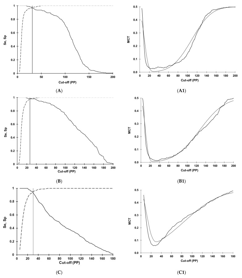
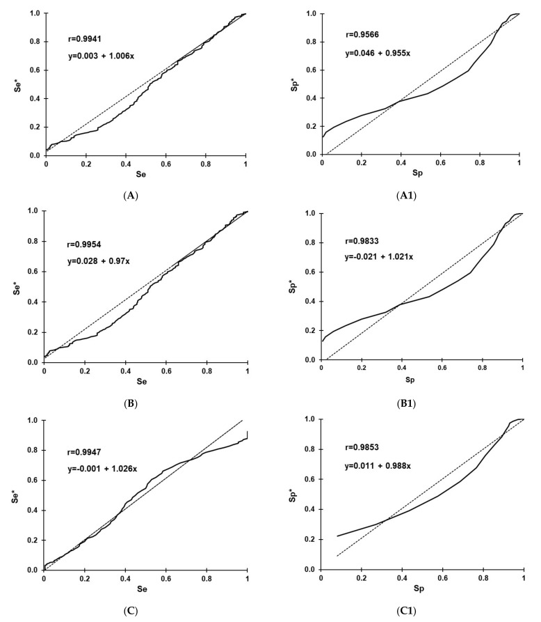
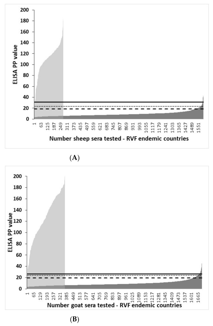
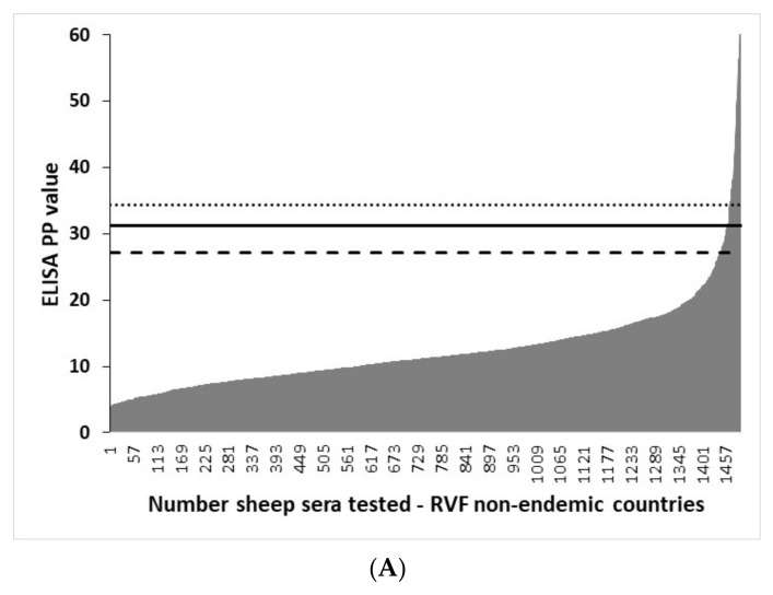
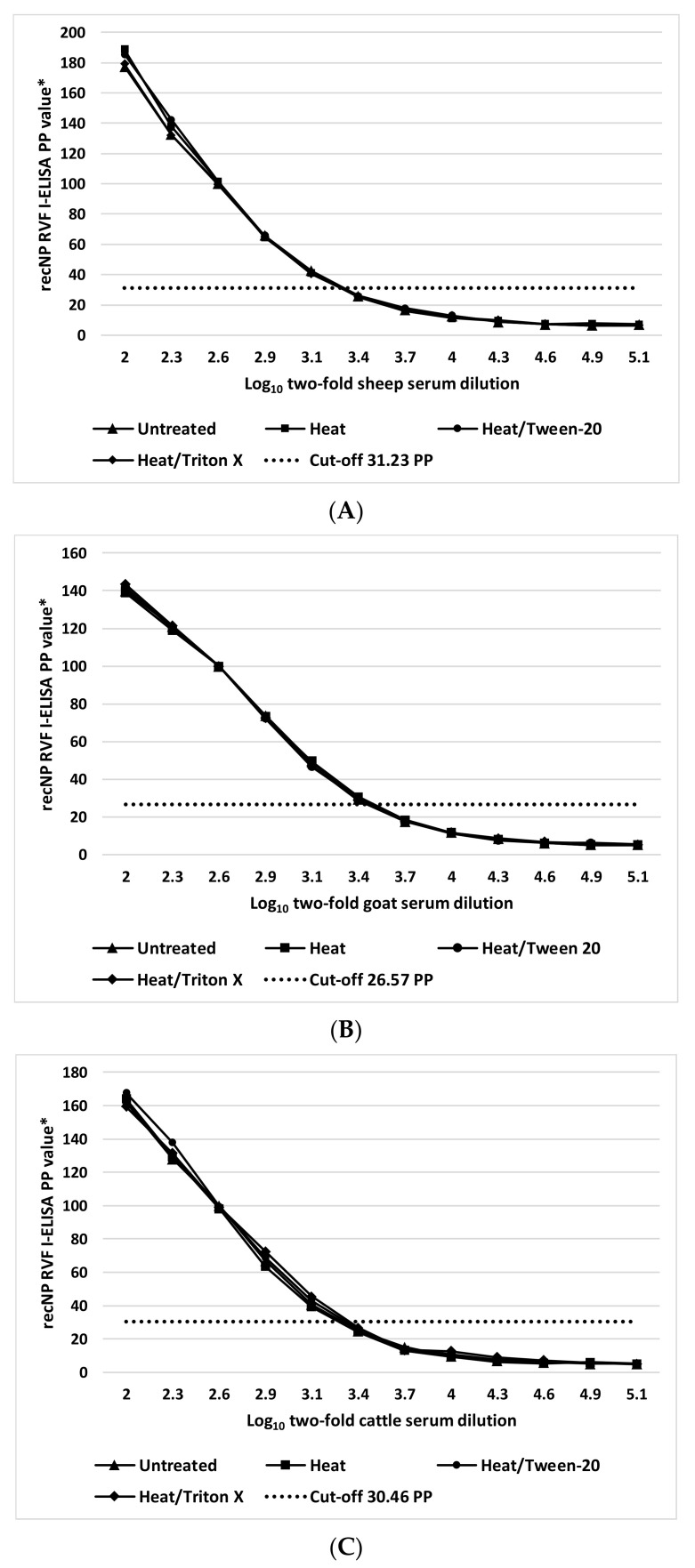
Diagnostic performance of an indirect enzyme-linked immunosorbent assay (I-ELISA) based on a recombinant nucleocapsid protein (rNP) of the Rift Valley fever virus (RVFV) was validated for the detection of the IgG antibody in sheep (n = 3367), goat (n = 2632), and cattle (n = 3819) sera. Validation data sets were dichotomized according to the results of a virus neutralization test in sera obtained from RVF-endemic (Burkina Faso, Democratic Republic of Congo, Mozambique, Senegal, Uganda, and Yemen) and RVF-free countries (France, Poland, and the USA). Cut-off values were defined using the two-graph receiver operating characteristic analysis. Estimates of the diagnostic specificity of the RVFV rNP I-ELISA in animals from RVF-endemic countries ranged from 98.6% (cattle) to 99.5% (sheep) while in those originating from RVF-free countries, they ranged from 97.7% (sheep) to 98.1% (goats). Estimates of the diagnostic sensitivity in ruminants from RVF-endemic countries ranged from 90.7% (cattle) to 100% (goats). The results of this large-scale international validation study demonstrate the high diagnostic accuracy of the RVFV rNP I-ELISA. Standard incubation and inactivation procedures evaluated did not have an adverse effect on the detectable levels of the anti-RVFV IgG in ruminant sera and thus, together with recombinant antigen-based I-ELISA, provide a simple, safe, and robust diagnostic platform that can be automated and carried out outside expensive bio-containment facilities. These advantages are particularly important for less-resourced countries where there is a need to accelerate and improve RVF surveillance and research on epidemiology as well as to advance disease control measures.

Keywords: IgG antibody; Rift Valley fever virus; diagnostic accuracy; domestic ruminants; enzyme-linked immunosorbent assay; recombinant nucleocapsid; validation.


Conflict of interest statement

The authors declare no conflict of interest. The funder had no role in the design of the study; the collection, analyses, or interpretation of data; in the writing of the manuscript; or the decision to publish the results.

Figures

Figure 1
Figure 1
Optimization of cut-offs for the Rift Valley fever IgG indirect ELISA based on recombinant nucleocapsid antigen in sheep (A), goats (B), and cattle (C) using the two-graph receiver operating characteristic analysis (TG-ROC). The insertion point of the sensitivity (Se, smooth line) and specificity (Sp, dashed line) graphs represents a cut-off PP value (31.23, 26.57, and 30.46, respectively) at which the highest and equivalent test parameters (Se = Sp) are achieved at 95% accuracy level. Using the misclassification cost term (MCT) option of the TG-ROC, at these cut-off values, the overall misclassification costs in sheep (A1), goats (B1), and cattle (C1) become minimal (0.0045, 0.0054, and 0.0625, respectively) under the assumption of 50% disease prevalence and equal costs of false-positive and false-negative results. The two MCT curves represent values based on non-parametric (smooth line) or parametric (dashed line) estimates of Se and Sp derived from datasets in field-collected sera.
Figure 2
Figure 2
Linear correlation analysis of the non-parametric (smooth line) sensitivity (Se) vs. parametric (dashed line) sensitivity (Se*) in sheep (A), goats (B), and cattle (C), and the non-parametric specificity (Sp) vs. parametric specificity (Sp*) in sheep (A1), goats (B1) and cattle (C1).
Figure 3
Figure 3
The effect of different I-ELISA cut-off values on the categorization between sera from RVF-endemic countries tested positive or negative in the virus neutralization test. Distribution of IgG I-ELISA PP values in sera tested positive in VNT (grey area): (A) sheep (n = 275), (B) goats (n = 369), and (C) cattle (n = 356). Distribution of IgG I-ELISA PP values in sera tested negative in VNT (dark area): (A) sheep (n = 1874), (B) goats (n = 2072), and (C) cattle (n = 2864) in the VNT. Sera ordered according to ELISA PP values. Vertical lines indicate the ELISA cut-off values determined by the TG-ROC analysis (solid line), and as mean plus three (dotted line) and two (slashed line) standard deviations observed in the VNT-negative sera.
Figure 3
Figure 3
The effect of different I-ELISA cut-off values on the categorization between sera from RVF-endemic countries tested positive or negative in the virus neutralization test. Distribution of IgG I-ELISA PP values in sera tested positive in VNT (grey area): (A) sheep (n = 275), (B) goats (n = 369), and (C) cattle (n = 356). Distribution of IgG I-ELISA PP values in sera tested negative in VNT (dark area): (A) sheep (n = 1874), (B) goats (n = 2072), and (C) cattle (n = 2864) in the VNT. Sera ordered according to ELISA PP values. Vertical lines indicate the ELISA cut-off values determined by the TG-ROC analysis (solid line), and as mean plus three (dotted line) and two (slashed line) standard deviations observed in the VNT-negative sera.
Figure 4
Figure 4
The effect of different I-ELISA cut-off values on the categorization of sera from RVF-non-endemic countries tested negative in the virus neutralization test. Distribution of IgG I-ELISA PP values (grey area) in (A) sheep (n = 1493), (B) goats (n = 560), and (C) cattle (n = 955). Sera ordered according to ELISA PP values. Vertical lines indicate the ELISA cut-off values determined by the TG-ROC analysis (solid line) in ruminant serum panels from RVF-endemic countries and as mean plus three (dotted line) and as mean plus two (slashed line) standard deviations observed in the VNT-negative serum panels from RVF-free countries.
Figure 4
Figure 4
The effect of different I-ELISA cut-off values on the categorization of sera from RVF-non-endemic countries tested negative in the virus neutralization test. Distribution of IgG I-ELISA PP values (grey area) in (A) sheep (n = 1493), (B) goats (n = 560), and (C) cattle (n = 955). Sera ordered according to ELISA PP values. Vertical lines indicate the ELISA cut-off values determined by the TG-ROC analysis (solid line) in ruminant serum panels from RVF-endemic countries and as mean plus three (dotted line) and as mean plus two (slashed line) standard deviations observed in the VNT-negative serum panels from RVF-free countries.
Figure 5
Figure 5
Dose-response kinetics of a positive RVFV IgG sheep (A), goat (B), and cattle (C) serum before and after different inactivation procedures measured by recombinant nucleocapsid I-ELISA. * Percent positivity of internal positive control serum.

References

    1. Bird B.H., Ksiazek T.G., Nichol S.T., Maclachlan N.J. Rift Valley fever virus. J. Am. Vet. Med. Assoc. 2009;234:883–893. doi: 10.2460/javma.234.7.883. - DOI - PubMed
    1. Chevalier V., Pepin M., Plee L., Lancelot R. Rift Valley fever—A threat for Europe? Eurosurveillance. 2010;15:19506. doi: 10.2807/ese.15.10.19506-en. - DOI - PubMed
    1. Paweska J.T., Jansen van Vuren P. Rift Valley fever virus: A virus with potential for global emergence. In: Johnson N., editor. The Role of Animals in Emerging Viral Diseases. Elsevier; Amsterdam, The Netherlands: Academic Press; Cambridge, MA, USA: 2013. pp. 169–200.
    1. McMillen C.M., Hartman A.L. Rift Valley fever in animals and humans: Current perspectives. Antivir. Res. 2018;156:29–37. doi: 10.1016/j.antiviral.2018.05.009. - DOI - PMC - PubMed
    1. Mehand M.S., Al-Shorbaji F., Millett P., Murgue B. The WHO R&D Blueprint: 2018 review of emerging infectious diseases requiring urgent research and development efforts. Antivir. Res. 2018;159:63–67. - PMC - PubMed

Publication types

MeSH terms

LinkOut - more resources