Review
. 2021 Aug 20;13(8):1657.
doi: 10.3390/v13081657.

Equine Influenza Virus and Vaccines

Affiliations
Review

Equine Influenza Virus and Vaccines

Fatai S Oladunni et al. Viruses. .

Abstract

Equine influenza virus (EIV) is a constantly evolving viral pathogen that is responsible for yearly outbreaks of respiratory disease in horses termed equine influenza (EI). There is currently no evidence of circulation of the original H7N7 strain of EIV worldwide; however, the EIV H3N8 strain, which was first isolated in the early 1960s, remains a major threat to most of the world's horse populations. It can also infect dogs. The ability of EIV to constantly accumulate mutations in its antibody-binding sites enables it to evade host protective immunity, making it a successful viral pathogen. Clinical and virological protection against EIV is achieved by stimulation of strong cellular and humoral immunity in vaccinated horses. However, despite EI vaccine updates over the years, EIV remains relevant, because the protective effects of vaccines decay and permit subclinical infections that facilitate transmission into susceptible populations. In this review, we describe how the evolution of EIV drives repeated EI outbreaks even in horse populations with supposedly high vaccination coverage. Next, we discuss the approaches employed to develop efficacious EI vaccines for commercial use and the existing system for recommendations on updating vaccines based on available clinical and virological data to improve protective immunity in vaccinated horse populations. Understanding how EIV biology can be better harnessed to improve EI vaccines is central to controlling EI.

Keywords: H3N8; adaptive immunity; cellular immunity; equine influenza; equine influenza vaccine; equine influenza virus; experimental infection; humoral immunity; surveillance.


Conflict of interest statement

The authors declare no conflict of interest.

Figures

Figure 1
Figure 1
Origin and mode of transmission of EIV: Aquatic birds serve as the natural reservoir of avian influenza, a progenitor of EIV. Following infection of a horse, the virus can be spread to other animals in aerosolized droplets by coughing or through fomite transmission. International transport of horses facilitates the spread of the virus to new geographical locations, especially during horse events such as racing or shows. Dogs can also become infected and transmit the virus to other dogs.
Figure 2
Figure 2
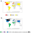
Incidence of EIV in the equine population over the last decade, compiled from OIE ESP on Equine Influenza Vaccine Composition reports, 2010 to 2019. (A) Countries are ranked based on the average of incidence rate, i.e., number of years reported to OIE ESP over a period of 10 years. (B) Clades responsible for these outbreaks indicated: An incidence rate >50% is classified as high, 50% is moderate, and <50% is low. NA, not available.
Figure 3
Figure 3
Schematic representation of an influenza A virus (IAV) genome organization and virion structure. (A) The genome organization of the eight negative-sense RNA gene segments (PB2, PB1, PA, HA, NP, NA, M, and NS) of IAV. All segments 1—8 (as in Table 2) are numbered according to the conventional representation from 3′ to 5′. The black boxes at the end of each viral segment indicate the noncoding regions (NCR). White boxes show the packaging signals present at the 3’ and 5′ ends of each gene segment. (B) The basic architecture of IAV contains envelope surface glycoproteins HA, NA, and M2 ion channel protein, all embedded within the host cell-derived lipid bilayer envelope. Immediately underneath the envelope is a well-organized layer of M1 and NEP proteins surrounding the eight negative-sense viral RNA segments.
Figure 4
Figure 4
Schematic of evolution of EIV HA, 1963 to 2021. The major lineages as discussed in the text are indicated, as are representative virus strains. Not to scale.
Figure 5
Figure 5
EI vaccine technologies: Each EI vaccine system differs in its antigenic composition and structural organization. These differences impact on the immunogenicity of the vaccine type.
Figure 6
Figure 6
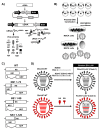
Generation of recombinant EIV LAIV using plasmid-based reverse genetic approaches: (A) Schematic illustration of the ambisense plasmids to generate recombinant EIV: Ambisense plasmids for the rescue of recombinant EIV contain the human polymerase I promoter (hPol-I, gray arrow) and the mouse polymerase I terminator (T, gray box) to regulate the expression of the negative-sense vRNAs. In the opposite direction to the Pol-I cassette, the polymerase II-dependent promoter (Pol-II, black arrow) and the bovine growth hormone polyadenylation sequence (BGH, black box) produce viral proteins. The vRNAs generated from the Pol-I cassette are recognized by the EIV polymerase subunits PB2, PB1, and PA that, together with the viral NP, form the viral ribonucleoprotein (vRNP) complexes involved in viral genome replication and gene transcription. Transcription of the vRNA results in viral mRNA and protein production. Replication of vRNAs results in complementary (c)RNAs for the amplification and synthesis of new vRNAs that are incorporated into nascent virions. (B) Generation of recombinant EIV using plasmid-based reverse genetics: Cocultures of human 293T and MDCK cells are cotransfected with the eight ambisense plasmids containing the EIV PB2, PB1, PA, HA, NP, NA, M, and NS segments. EIV generated from transfected cells is amplified in fresh monolayers of MDCK cells or in chicken embryonated eggs. (C) LAIV based on truncations of EIV NS1 protein: Schematic representation of WT and 1–73, 1–99, and 1–126 NS1 truncated NS viral segments. Black lines represent stop codons introduced to truncate NS1. Lines at the end of the NS segment indicate the 3’and 5’ NCR. (D) Schematic representation for the development of an EIV bivalent LAIV: The mutations responsible of the ts, ca, and att phenotype of the human A/Ann Arbor/6/60 H2N2 MDV LAIV were introduced into the PB2 (N265S) and PB1 (K391E, E581G and A661T) viral segments of A/equine/Ohio/1/2003 H3N8 WT (Ohio/03 WT) to generate the FC-1 EIV monovalent LAIV (Ohio/03 LAIV). The backbone of Ohio/03 LAIV was used as an MDV to generate a recombinant virus containing the HA and NA glycoproteins of the FC-2 A/Richmond/1/2007 H3N8 WT (Rich/07 WT) to develop the FC-2 EIV monovalent LAIV (Rich/07 LAIV). The bivalent EIV LAIV was developed by combining the monovalent Ohio/03 and Rich/07 LAIVs.

References

    1. Timoney P.J. Equine Infectious Diseases. 2nd ed. Elsevier; Amsterdam, The Netherlands: 2014. Infectious diseases and international movement of horses; pp. 544–551.e1.
    1. Dominguez M., Munstermann S., de Guindos I., Timoney P. Equine disease events resulting from international horse movements: Systematic review and lessons learned. Equine. Vet. J. 2016;48:641–653. doi: 10.1111/evj.12523. - DOI - PubMed
    1. Guthrie A.J., Stevens K.B., Bosman P.P. The circumstances surrounding the outbreak and spread of equine influenza in South Africa. Rev. Sci. Tech. 1999;18:179–185. doi: 10.20506/rst.18.1.1155. - DOI - PubMed
    1. Kawaoka Y., Bean W.J., Webster R.G. Evolution of the hemagglutinin of equine H3 influenza viruses. Virology. 1989;169:283–292. doi: 10.1016/0042-6822(89)90153-0. - DOI - PubMed
    1. Kawaoka Y., Webster R.G. Origin of the hemagglutinin on A/Equine/Johannesburg/86 (H3N8): The first known equine influenza outbreak in South Africa. Arch. Virol. 1989;106:159–164. doi: 10.1007/BF01311048. - DOI - PubMed

Publication types

MeSH terms