

Acute Disseminated Encephalomyelitis and Acute Hemorrhagic Leukoencephalitis Following COVID-19: Systematic Review and Meta-synthesis
- PMID: 34452974
- PMCID: PMC8404207
- DOI: 10.1212/NXI.0000000000001080
Acute Disseminated Encephalomyelitis and Acute Hemorrhagic Leukoencephalitis Following COVID-19: Systematic Review and Meta-synthesis
Abstract
Background and objectives: Since the onset of the COVID-19 pandemic, a growing number of reports have described cases of acute disseminated encephalomyelitis (ADEM) and acute hemorrhagic leukoencephalitis (AHLE) following infection with COVID-19. Given their relatively rare occurrence, the primary objective of this systematic review was to synthesize their clinical features, response to treatments, and clinical outcomes to better understand the nature of this neurologic consequence of COVID-19 infection.
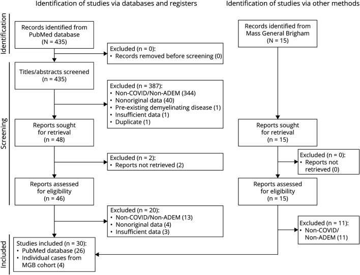
Methods: Patients with a history of COVID-19 infection were included if their reports provided adequate detail to confirm a diagnosis of ADEM or AHLE by virtue of clinical features, radiographic abnormalities, and histopathologic findings. Cases purported to be secondary to vaccination against COVID-19 or occurring in the context of a preexisting relapsing CNS demyelinating disease were excluded. Case reports and series were identified via PubMed on May 17, 2021, and 4 additional cases from the authors' hospital files supplemented the systematic review of the literature. Summary statistics were used to describe variables using a complete case analysis approach.
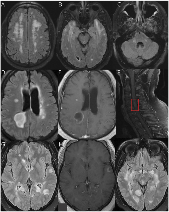
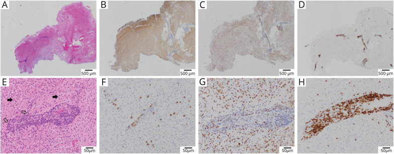
Results: Forty-six patients (28 men, median age 49.5 years, 1/3 >50 years old) were analyzed, derived from 26 case reports or series originating from 8 countries alongside 4 patient cases from the authors' hospital files. COVID-19 infection was laboratory confirmed in 91% of cases, and infection severity necessitated intensive care in 67%. ADEM occurred in 31 cases, whereas AHLE occurred in 15, with a median presenting nadir modified Rankin Scale score of 5 (bedridden). Anti-MOG seropositivity was rare (1/15 patients tested). Noninflammatory CSF was present in 30%. Hemorrhage on brain MRI was identified in 42%. Seventy percent received immunomodulatory treatments, most commonly steroids, IV immunoglobulins, or plasmapheresis. The final mRS score was ≥4 in 64% of patients with adequate follow-up information, including 32% who died.
Discussion: In contrast to ADEM cases from the prepandemic era, reported post-COVID-19 ADEM and AHLE cases were often advanced in age at onset, experienced severe antecedent infection, displayed an unusually high rate of hemorrhage on neuroimaging, and routinely had poor neurologic outcomes, including a high mortality rate. Findings are limited by nonstandardized reporting of cases, truncated follow-up information, and presumed publication bias.
Copyright © 2021 The Author(s). Published by Wolters Kluwer Health, Inc. on behalf of the American Academy of Neurology.
Figures
References
-
- Krupp LB, Tardieu M, Amato MP, et al. International Pediatric Multiple Sclerosis Study Group criteria for pediatric multiple sclerosis and immune-mediated central nervous system demyelinating disorders: revisions to the 2007 definitions. Mult Scler. 2013;19(10):1261-1267. - PubMed
-
- Koelman DLH, Chahin S, Mar SS, et al. Acute disseminated encephalomyelitis in 228 patients: a retrospective, multicenter US study. Neurology. 2016;86(22):2085-2093. - PubMed
Publication types
MeSH terms
Substances
LinkOut - more resources
Full Text Sources
Medical