. 2021 Aug 31;118(35):e2101657118.
doi: 10.1073/pnas.2101657118.

Dual-mechanism estrogen receptor inhibitors

Affiliations

Dual-mechanism estrogen receptor inhibitors

Jian Min et al. Proc Natl Acad Sci U S A. .

Abstract

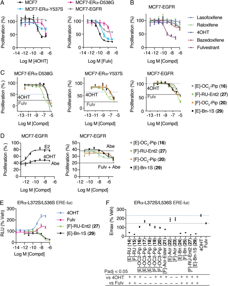
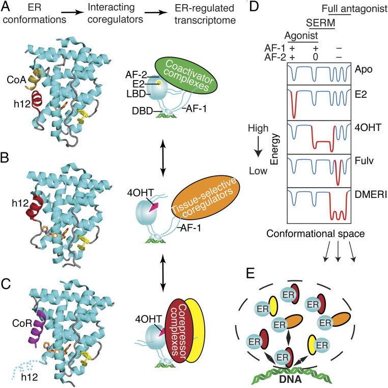
Efforts to improve estrogen receptor-α (ER)-targeted therapies in breast cancer have relied upon a single mechanism, with ligands having a single side chain on the ligand core that extends outward to determine antagonism of breast cancer growth. Here, we describe inhibitors with two ER-targeting moieties, one of which uses an alternate structural mechanism to generate full antagonism, freeing the side chain to independently determine other critical properties of the ligands. By combining two molecular targeting approaches into a single ER ligand, we have generated antiestrogens that function through new mechanisms and structural paradigms to achieve antagonism. These dual-mechanism ER inhibitors (DMERIs) cause alternate, noncanonical structural perturbations of the receptor ligand-binding domain (LBD) to antagonize proliferation in ER-positive breast cancer cells and in allele-specific resistance models. Our structural analyses with DMERIs highlight marked differences from current standard-of-care, single-mechanism antiestrogens. These findings uncover an enhanced flexibility of the ER LBD through which it can access nonconsensus conformational modes in response to DMERI binding, broadly and effectively suppressing ER activity.

Keywords: SERM; X-ray crystallography; breast cancer; cancer therapy; estrogen receptor.


Conflict of interest statement

Competing interest statement: J.A.K. is a founder and stockholder of Radius Health Inc. and a consultant of Celcuity Inc. B.S.K. is a consultant of Celcuity Inc.

Figures

Fig. 1.
Fig. 1.
Dual-mechanism ER inhibitors fully suppress breast cancer cell proliferation. (A) Chemical structure of the OBHS-N scaffold and the orientation of substituents R1 and R2, with respect to h11 and h12 in the ER LBD (when R1 has a substituent, R2 is −OCH3 group; when R2 has a substituent, R1 is −OH for compounds 14–29; SI Appendix, Fig. S2 A–H). (B) Proliferation of MCF-7 cells treated for 5 d with 4OHT, fulvestrant (Fulv), or the indicated compounds. Datapoints are mean ± SEM, N = 6. M, Molarity. The horizontal lines indicate the Emax for 4OHT and fulvestrant (Fulv). (C) The structure of [E]-OC2-Pip (16)-bound ER LBD showed that the E-ring substituted piperidine H-bonding to Asp351 in helix 3 (h3), while the F-ring shifts helix 11 (h11) by 2.4 Å compared to an agonist bound structure. 2Fo-Fc electron density map contoured to 0.9 σ within 2 Å of the ligand. (D) The structure of [F]-OC2-Pip (19)–bound ER LBD shows that its [F]-OC2-Pip side chain exits the ligand-binding pocket between h8 and h11, H-bonds to His524, and shifts h11 toward h12. (E) Summary of dose–response curves for compound inhibition of proliferation of MCF-7 cells, shown in SI Appendix, Fig. S2 A–H. Datapoints are mean ± SEM, n = 6. * indicates pAdj < 0.05 (one-way ANOVA) for compounds with Emax > fulvestrant. (F) Selected dose curves from E. The lines indicate the Emax for 4OHT and Fulv.
Fig. 2.
Fig. 2.
Dual-mechanism inhibitors destabilize helix 12 of ERα. (A) Structure of the ER LBD bound to [F]-OC3-Pip (20). 2Fo-Fc electron density map contoured at 1.0 σ shows the 20 F-ring facing outward between h3 and 11 toward h12, shifting h11 1.6 Å compared to an agonist bound structure. The 2Fo-Fc electron density map is contoured at 0.9 σ within 2 Å of the ligand. (B) The structure of ER with 20 shows that 20 is stabilized by contacts with Trp383, which stabilizes the altered conformer of h12 by contacting L539. (C) The structures of the ER LBD bound to 20 (coral) or raloxifene (gray) were superimposed, showing the 2.6-Å shift of h12 to contact the piperidine ring of 20. (D) Structure of the ER LBD with [E]-Bn-1S (29) shows that h12 could not be modeled in two of four subunits due to poor electron density. The A chain of h3 (yellow) is shown with the B chain superimposed (gray) to show the expected location of h12, which was not modeled. The 2Fo-Fc electron density map is contoured at 1.0 σ. (E) Structure of [F]-AcrEster (21)-bound ER showing different ligand-binding positions in the dimeric subunits. The A and B chains were superimposed and colored blue or coral. (F and G) Changes in ER-Y537S H/D exchange compared to the apo receptor. ER-Y537S LBD was incubated with the indicated ligands and then assayed for exchange of amide hydrogens with deuterium over time, as measured by mass spectrometry. Regions colored black were not detected (SI Appendix, Fig. S4).
Fig. 3.
Fig. 3.
SERM and SERD properties of DMERI ligands. (A) ER and β-actin levels in MCF-7 cells treated with the indicated compounds for 24 h. Whole-cell lysates were analyzed by Western blot. See also SI Appendix, Fig. S5 A and B. (B) Summary of dose–response curves of HepG2 cells transfected with 3xERE-luciferase reporter and treated with the indicated ligands. Datapoints are mean ± SEM, N= 3. *Significantly different from fulvestrant by one-way ANOVA, Sidak’s test adjusted P value (padj) < 0.05. n = 3 to 6. Dose curves are shown in SI Appendix, Fig. S5C.
Fig. 4.
Fig. 4.
Dual-mechanism inhibitors promote conformations of ER that are distinct from traditional single mechanism inhibitors. (A) Hierarchical clustering of MARCoNI FRET assay for interaction of full-length and WT ER with 154 peptides derived from nuclear receptor-interacting proteins and the indicated ligands. (B and C) MARCoNI Pearson correlations for 4OHT versus the indicated ligands. Fulvestrant (Fulv), GDC-0810 (GDC), or AZD9496 (AZD). r = Pearson correlation ligand versus 4OHT. (D) Hierarchical clustering of MARCONI data with ER-Y537S and the indicated ligands. (E) MARCoNI Pearson correlations for 4OHT versus [E]-C2-Pip (16) with the ER-Y537S. r = Pearson correlation (SI Appendix, Fig. S7).
Fig. 5.
Fig. 5.
Activity of ligands in allele-specific models of tamoxifen resistance. (AC) WT MCF7 cells and MCF7 cells engineered to express the mutant ERα-Y537S or ERα-D538G or overexpress EGFR were treated with the indicated SERM (16, 20) or SERD (27, 29) for 5 d and analyzed for inhibition of cell proliferation. Fulvestrant (Fulv). n = 3. Dashed lines indicate Emax values for 4OHT or fulvestrant (SI Appendix, Fig. S8). (D) MCF7-EGFR cells were treated with the indicated 1μM abemaciclib (Abe) and the indicated ligands for 5 d and analyzed for inhibition of cell proliferation. Fulvestrant (Fulv). n = 3. Dashed lines indicate Emax values for abemaciclib alone or fulvestrant (+Abe). (E and F) Structure-based model of tamoxifen and fulvestrant resistance. HepG2 liver cells were treated for 24 h with the indicated ligands. n = 6, except for 4OHT and Fulvestrant where n = 18. Data were analyzed by one-way ANOVA. Data are mean ± SEM.
Fig. 6.
Fig. 6.
Ligand-dependent control of ERα-LBD conformation and of ER coregulator recruitment and selection of activity states. (A) The active LBD conformation (7). (Left) Ribbon diagram of the ER LBD bound to estradiol. Helix 12 (h12, colored red) forms one side of the coactivator binding site, shown here binding to a peptide from Steroid Receptor Coactivator-2 (CoA colored yellow) from PDB entry 3UUD. (Right) Schematic of ERα bound to estradiol (E2), DNA, and a coactivator complex. With full agonists, the coactivator recruitment to the LBD surface, AF-2 nucleates binding of multiprotein coactivator complexes to other domains including AF-1 (Activation Function-1). DBD, DNA binding domain. Steroid Receptor Coactivators (SRCs) 1–3 bind to both AF-1 and AF-2 through separate interactions. (B) The inactive LBD conformer (7, 27). Left, Ribbon diagram of the ER LBD bound to an antagonist. Antagonists can flip h12 (colored red) into the coactivator/corepressor binding site, rendering the LBD inactive by blocking both coactivator and corepressor binding to AF-2, from PDB entry 2QXS. (Right) When h12 blocks both coactivators and corepressors from binding the LBD, the activity of AF-1 is cell-type specific. (C) The transcriptionally repressive LBD conformation (9, 12, 25, 53). (Left) Ribbon diagram of the ER LBD bound to a corepressor peptide, colored violet. When h12 is disordered by an antagonist, the LBD can bind an extended peptide motif found in transcriptional corepressors (8) from PDB entry 2JFA. (Right) Cartoon of ERα bound to 4OHT and a corepressor complex, repressing both AF-1 and AF-2 activity and mediating mediate chromatin compaction and inhibition of proliferative gene expression. (D) Energy diagram illustrating how ER ligands differ in stabilizing, specific, low-energy receptor conformations associated with transcriptional activity (+), inactivity (0), or repression (−) that are being driven by the activity state of AF-2 or AF-1. The dips in the curves represent different LBD conformations associated with the three AF-1/AF-2 activity states shown at the top, leftmost being the active state (a), the rightmost representing substates of the repressive state (b), and the middle the inactive state (c). When a state is stabilized by a particular type of ligand, the curves become deeper, with gray changed to red; the barrier heights between states indicate the ease of dynamic interchange among the states or substates. The DMERI showed multiple mechanisms of antagonism, represented by the multiple-favored repressor substates with reduced exchange barriers. (E) Dashed line indicates a transcriptional phase condensate with multiple receptor–coregulator complexes exchanging at an ER binding site, enabling multiple mechanisms of antagonism.

References

    1. Rugo H. S., et al. ., Endocrine therapy for hormone receptor-positive metastatic breast cancer: American Society of Clinical Oncology guideline. J. Clin. Oncol. 34, 3069–3103 (2016). - PubMed
    1. Tryfonidis K., Zardavas D., Katzenellenbogen B. S., Piccart M., Endocrine treatment in breast cancer: Cure, resistance and beyond. Cancer Treat. Rev. 50, 68–81 (2016). - PubMed
    1. Clarke R., Tyson J. J., Dixon J. M., Endocrine resistance in breast cancer–An overview and update. Mol. Cell. Endocrinol. 418, 220–234 (2015). - PMC - PubMed
    1. Lerner H. J., Band P. R., Israel L., Leung B. S., Phase II study of tamoxifen: Report of 74 patients with stage IV breast cancer. Cancer Treat. Rep. 60, 1431–1435 (1976). - PubMed
    1. Jordan V. C., Phelps E., Lindgren J. U., Effects of anti-estrogens on bone in castrated and intact female rats. Breast Cancer Res. Treat. 10, 31–35 (1987). - PubMed

Publication types