. 2021 Aug 31;118(35):e2105961118.
doi: 10.1073/pnas.2105961118.

Structural basis for the activation of the DEAD-box RNA helicase DbpA by the nascent ribosome

Affiliations

Structural basis for the activation of the DEAD-box RNA helicase DbpA by the nascent ribosome

Jan Philip Wurm et al. Proc Natl Acad Sci U S A. .

Abstract

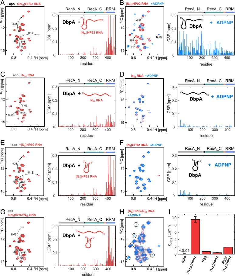
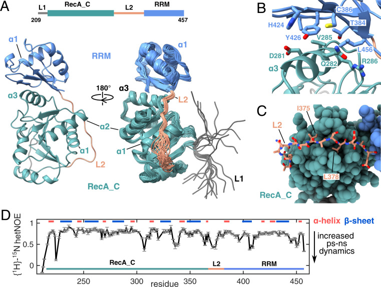
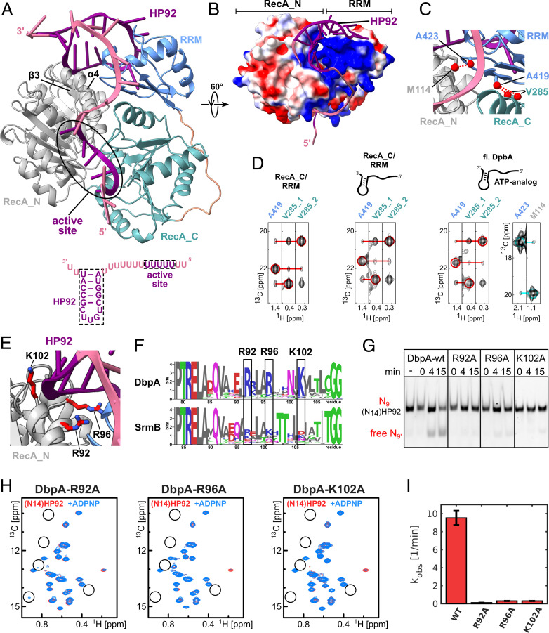
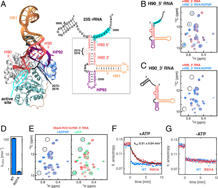
The adenosine triphosphate (ATP)-dependent DEAD-box RNA helicase DbpA from Escherichia coli functions in ribosome biogenesis. DbpA is targeted to the nascent 50S subunit by an ancillary, carboxyl-terminal RNA recognition motif (RRM) that specifically binds to hairpin 92 (HP92) of the 23S ribosomal RNA (rRNA). The interaction between HP92 and the RRM is required for the helicase activity of the RecA-like core domains of DbpA. Here, we elucidate the structural basis by which DbpA activity is endorsed when the enzyme interacts with the maturing ribosome. We used nuclear magnetic resonance (NMR) spectroscopy to show that the RRM and the carboxyl-terminal RecA-like domain tightly interact. This orients HP92 such that this RNA hairpin can form electrostatic interactions with a positively charged patch in the N-terminal RecA-like domain. Consequently, the enzyme can stably adopt the catalytically important, closed conformation. The substrate binding mode in this complex reveals that a region 5' to helix 90 in the maturing ribosome is specifically targeted by DbpA. Finally, our results indicate that the ribosome maturation defects induced by a dominant negative DbpA mutation are caused by a delayed dissociation of DbpA from the nascent ribosome. Taken together, our findings provide unique insights into the important regulatory mechanism that modulates the activity of DbpA.

Keywords: DEAD-box helicase; NMR spectroscopy; enzyme regulation; molecular mechanism; ribosome assembly.


Conflict of interest statement

The authors declare no competing interest.

Figures

Fig. 1.
Fig. 1.
DbpA domain organization, rRNA binding, and activation of DbpA activity by HP92. (A) Domain organization of E. coli DbpA. The linker L1 and L2 that connect the three domains are shown in black and light brown, respectively. Residue numbers at the domain boundaries are indicated. (B) Secondary structure of the 23S rRNA (nt 2,505 to 2,584) as found in the mature 50S subunit. HP92, which is recognized specifically by the DbpA RRM, is shown in violet. (C) Helicase assays that show that the binding of HP92 to the RRM in trans enables duplex unwinding by DbpA. Unwinding of an RNA duplex consisting of a fluorescently labeled 9mer RNA (red) and the unstructured N32 RNA (Bottom) is followed by native polyacrylamide gel electrophoresis (PAGE) analysis. The nonfluorescently labeled HP92–derived RNAs that were added in trans are indicated on the Top. No unwinding is observed in the absence of HP92 (−) or in the presence of a short, stabilized HP92 fragment without ssRNA overhangs (S). HP92 constructs containing 5′ overhangs of 4 to 12 nt enable duplex unwinding by DbpA.
Fig. 2.
Fig. 2.
HP92 is required for the formation of the catalytically important closed conformation of DbpA. (AF) Titrations of NMR-active (ILMVA-labeled) full-length DbpA with different NMR-inactive RNA constructs and the ATP analog ADPNP: (Left) Isoleucine region of the methyl TROSY spectra of free DbpA (black), after addition of different RNA constructs (red), or after addition of different RNA constructs and ADPNP (blue). (Right) CSPs of the ILMVA methyl groups caused by binding of RNA to DbpA (A, C, and E) or by binding of ADPNP to the DbpA/RNA complex (B, D, and F) are plotted against the residue number. DbpA domains are indicated on top. (A) An RNA that contains a single-stranded region of 14 nt plus HP92 [(N14)HP92 RNA] binds mainly to the RRM. The signals of three Ile Cδ1 methyl groups of the RRM are labeled. (B) Addition of ADPNP to the DbpA/ (N14)HP92 RNA complex induces strong CSPs in both core domains and leads to the formation of the closed helicase conformation. Assignments of several Ile Cδ1 signals are indicated in the spectrum. (C) An unstructured RNA of 32 nt (N32) that lacks HP92 shows weak binding to the RRM. (D) For the unstructured N32 RNA, the helicase does not form the closed conformation upon addition of ADPNP, and only small CSPs in the vicinity of the ATP binding site are observed. (E) The (N2)HP92 RNA that contains HP92 but that lacks the 5′ ss overhang binds to the RRM similar to the (N14)HP92 RNA (compare to A). (F) Addition of ADPNP to the DbpA/(N2)HP92 RNA complex does not result in the formation of the closed helicase conformation and CSPs are only observed in the vicinity of the ATP binding site. (G) Simultaneous addition of the (N2)HP92 and the N32 RNAs leads to similar CSPs in the RRM as are observed for the interaction with the (N2)HP92 RNA (compare to E), although several of the shifted signals are broadened. (H) Addition of ADPPNP to this DbpA/RNA complex causes the partial (∼10 to 15%) formation of the closed conformation. This implies that the helicase can adopt the closed conformation on a ssRNA when HP92 is bound in trans. The characteristic Ile Cδ1 signals of the closed state are indicated by circles (compare to B). (I) Stimulation of the ATPase activity of DbpA by the RNAs that were used in the NMR experiments (AH). ATP turnover rates were determined using a coupled pyruvate kinase–lactate dehydrogenase assay. The ATPase activity in the absence of RNA is below the detection limit of the assay. The ATPase activity of DbpA thus correlates with the degree to which the closed conformation is adopted.
Fig. 3.
Fig. 3.
Solution structure of the DbpA RecA_C/RRM domains. (A) The construct used for the NMR structure determination is shown on top and includes the RecA_C domain and the RRM of DbpA. A cartoon representation of the structure with the lowest target function is shown on the left. The bundle of 20 NMR structures is shown on the right, rotated by 90°. The structure reveals that the RRM (light blue) tightly interacts with α-3 of the RecA_C domain (green) via its beta sheet surface. Note that the interdomain orientation is well defined. L1 (gray) and the N-terminal half of L2 (light brown) are less well defined due to increased local dynamics (see below). (B) Closeup of the interface between the RecA_C and RRM domains. Central residues are labeled and shown in stick representation. (C) Closeup of the interaction between the carboxyl-terminal part of L2 and the RecA_C domain. I375 and I378 of L2 that form hydrophobic interactions with the RecA_C domain are labeled. (D) {1H}-15N hetNOE values of the RecA_C/RRM construct that report on fast internal protein motions. Decreased values in several loop regions including the N-terminal half of L1 reveal increased dynamics on the ps to ns timescale. Secondary structure elements are indicated on top.
Fig. 4.
Fig. 4.
A model of the closed state of DbpA reveals stabilizing interactions between the RecA_N domain and HP92 that enable helicase activity. (A) The model of the closed state based on the solution structure of the RecA_C/RRM construct shows that HP92 and the RRM contact the RecA_N domain in the closed state. HP92 and 5 nt of the 5′ ss overhang bound to the ssRNA binding site of the helicase core were modeled based on previously published structures (3, 17). These regions are shown in purple, and the rest of the RNA is shown in pale violet. RRM, RecA_N and RecA_C domains are shown in blue, gray, and green, respectively. β-3 and α-4 of the RecA_N domain that interact with HP92 and the RRM are indicated. The RNA construct that is bound to the helicase in the model is shown on the bottom. (B) Surface representation the modeled DbpA colored according to the electrostatic surface potential (positive, blue; negative, red). The structure is rotated by 60° compared to A. The stem of HP92 packs into a positively charged groove formed between RRM and RecA_N domains. (C) Closeup of the interdomain interface of the closed state. Methyl groups that are predicted to show strong interdomain NOEs (dashed, red lines) are shown in red. The short distance between V285 and A419 (Right) is used to validate the orientation between the RecA_C and RRM domains. The short distance between A423 and M114 reports on the correct orientation between the RecA_N and RRM domains. (D) CCH-NOESY strips along the 13C-NOE dimension that show the interdomain NOEs for the RecA_C/RRM construct in the free form (Left), bound to (N14)HP92 RNA (Middle), or for full length DbpA in the closed state (Right). Interdomain NOEs between the RecA_C (V285) and RRM domains (A419) are indicated by red lines. The interdomain NOE between the RecA_N and RRM domains in the closed state (between A423 and M114) is indicated by a cyan line. Diagonal peaks are indicated by circles. (E) Closeup of the interaction between the RecA_N domain and HP92 in the closed state. The conserved Arg/Lys residues (R92/R96/K102) are shown in stick representation (red). (F) Sequence logos for DbpA (Top) and the unrelated DEAD-box helicase SrmB (Bottom). Residues R92, R96, and K102 are highly conserved across DbpA homologs, whereas the equivalent positions in SrmB are not conserved. The PTRELAXQ (Left) and GG motifs (Right) belong to the characteristic sequence motifs of DEAD-box helicases. (G) Mutation of the conserved Arg/Lys residues abolishes the helicase activity of DbpA. Unwinding of a fluorescently labeled 9mer RNA hybridized to (N14)HP92 RNA is followed by native PAGE after 0-, 4-, and 15-min incubation time. DbpA-wt shows robust unwinding after 15-min incubation time; no unwinding is observed after 15 min for the wt in the absence of ATP (−). Neither of the DbpA-R92A, -R96A, and -K102A mutants shows significant unwinding after 15 min. (H) Ile region of the methyl TROSY spectra of the DbpA-R92A, -R96A, and -K102A mutants bound to (N14)HP92 RNA (red) and after addition of ADPNP (blue). The positions of some of the characteristic signals of the closed state are indicated by circles. The mutations impair the formation of the closed conformation (R96A and K102A) or strongly reduce the population of the closed conformation (R92A) (compare to Fig. 2B). (I) Comparison of the ATPase activity of DbpA-wt with the -R92A, -R96A, and -K102A mutants. All measurements were performed in the presence of (N14)HP92 RNA. The mutations strongly reduce the ATPase activity of DbpA.
Fig. 5.
Fig. 5.
Interaction of DbpA with its target sequence in the 23S rRNA and effects of the DbpA-R331A. (A) Model of DbpA in the closed state bound to HP92 in the context of the 23S rRNA [nt 2,508 to 2,581, based on the YxiN RRM/23S rRNA structure (17)] (Left) and secondary structure of the 23S rRNA fragment (nt 2,496 to 2,604) (Right). The part of the rRNA that is observed in the structure is marked by a dashed box, and the secondary structure is depicted according to the crystal structure of the YxiN RRM/23S rRNA complex. The 5′ and 3′ parts of H90 (red) are indicated. The 5′ part of H90 points toward the ssRNA binding site of the helicase core. This places the region 5′ to H90 (nt 2,501 to 2,508, highlighted in cyan) in the correct orientation to interact with the active site (the ssRNA binding site and its 5′ to 3′ directionality are indicated in cyan). Note that nt 2,572 to 2,576 are not resolved in the crystal structure. (B) Secondary structure of the H90_5′ RNA lacking the ss region 3′ to H90 (Left) and Ile region of the methyl TROSY spectra of free DbpA (black), after addition of H90_5′ RNA (red) (Middle) or bound to H90_5′ RNA and ADPNP (blue) (Right). The RNA region that is proposed to interact with the helicase ssRNA binding site is highlighted in cyan. Characteristic signals of the closed state are marked by circles. (C) Same as B but for the H90_3′ RNA, which contains a ssRNA region 3′ to H90. No closed state is formed in the presence of H90_3′ RNA and ADPPNP, indicating that the ssRNA region 3′ to H90 is not a substrate region for DbpA. (D) ATP turnover of DbpA-wt and -R331A mutant in the presence of the H90_5′ RNA. The R331A mutation strongly reduces the ATPase activity of DbpA. (E) Ile region of the methyl TROSY spectra of DbpA-R331A bound to H90_5′ RNA (red) and after addition of ADPNP (Left, blue) or ATP (Right, green). ADPNP induces the formation of a very small population of the closed conformation (note the weak signal at the bottom left), whereas ATP leads to complete conversion to the closed conformation. Characteristic signals of the closed state are marked by circles. (F and G) Fluorescence anisotropy time traces of fluorescently labeled H90_5′ RNA in complex with DbpA in the presence (F) or absence (G) of ATP. At −2 min, the complex between labeled H90_5′ RNA and DbpA was formed (anisotropy ∼0.14). At t = 0, a 100-fold excess of unlabeled H90_5′ RNA was added. Dissociation of the labeled H90_5′ RNA leads to a decrease in fluorescence anisotropy. For DbpA-wt, dissociation is essentially complete after the dead time of the experiment (10 s, indicated by the vertical gray bar). The dissociation rate of the complex is decreased to 0.51 min−1 for the DbpA-R331A mutant.

Similar articles

Cited by

References

    1. Linder P., Jankowsky E., From unwinding to clamping - The DEAD box RNA helicase family. Nat. Rev. Mol. Cell Biol. 12, 505–516 (2011). - PubMed
    1. Putnam A. A., Jankowsky E., DEAD-box helicases as integrators of RNA, nucleotide and protein binding. Biochim. Biophys. Acta 1829, 884–893 (2013). - PMC - PubMed
    1. Sengoku T., Nureki O., Nakamura A., Kobayashi S., Yokoyama S., Structural basis for RNA unwinding by the DEAD-box protein Drosophila Vasa. Cell 125, 287–300 (2006). - PubMed
    1. Theissen B., Karow A. R., Köhler J., Gubaev A., Klostermeier D., Cooperative binding of ATP and RNA induces a closed conformation in a DEAD box RNA helicase. Proc. Natl. Acad. Sci. U.S.A. 105, 548–553 (2008). - PMC - PubMed
    1. Henn A., Bradley M. J., De La Cruz E. M., ATP utilization and RNA conformational rearrangement by DEAD-box proteins. Annu. Rev. Biophys. 41, 247–267 (2012). - PMC - PubMed

Publication types

MeSH terms

LinkOut - more resources