. 2021 Aug 27;12(1):5166.
doi: 10.1038/s41467-021-25429-2.

The structure of the bacterial DNA segregation ATPase filament reveals the conformational plasticity of ParA upon DNA binding

Affiliations

The structure of the bacterial DNA segregation ATPase filament reveals the conformational plasticity of ParA upon DNA binding

Alexandra V Parker et al. Nat Commun. .

Abstract

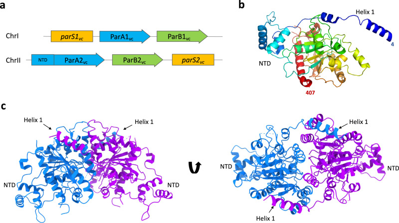
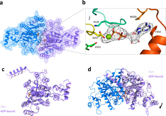
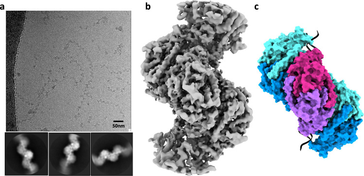
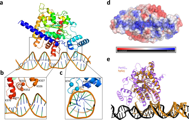
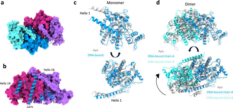
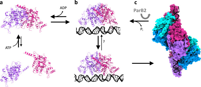
The efficient segregation of replicated genetic material is an essential step for cell division. Bacterial cells use several evolutionarily-distinct genome segregation systems, the most common of which is the type I Par system. It consists of an adapter protein, ParB, that binds to the DNA cargo via interaction with the parS DNA sequence; and an ATPase, ParA, that binds nonspecific DNA and mediates cargo transport. However, the molecular details of how this system functions are not well understood. Here, we report the cryo-EM structure of the Vibrio cholerae ParA2 filament bound to DNA, as well as the crystal structures of this protein in various nucleotide states. These structures show that ParA forms a left-handed filament on DNA, stabilized by nucleotide binding, and that ParA undergoes profound structural rearrangements upon DNA binding and filament assembly. Collectively, our data suggest the structural basis for ParA's cooperative binding to DNA and the formation of high ParA density regions on the nucleoid.


Conflict of interest statement

The authors declare no competing interests.

Figures

Fig. 1
Fig. 1. Structure of the ParA2vc dimer.
a Schematic representation of the chromosome segregation systems for the V. cholerae chromosome 1 (top), and chromosome II (bottom). The centromere-like partition site is in yellow (parS), the adapter parS binding protein is in green (ParB) and the NTPase protein is in blue (ParA), with the additional N-terminal domain (NTD) found only in ChrII shown. b Cartoon representation of the ParA2vc crystal structure, in rainbow coloring, starting from blue at the N terminus, to red at the C terminus. c The crystallographic dimer of ParA2vc shown from the side (left) and top (right), with the two symmetry-related pairs in blue and magenta, respectively. The NTD and helix 1 are indicated.
Fig. 2
Fig. 2. Structure of ParA2vc bound to ADP.
a Surface representation of the crystal structure of a ParA2vc-ADP dimer, coloured as in Fig. 1c. The ADP and Mg molecules, present in each ParA2vc molecules are shown in sticks and sphere representations, respectively. b Close-up view of the ADP and Mg in one of the ParA2 molecules. The composite omit map is shown around the nucleotide. Residues that interact with the nucleotide and Mg are shown in sticks. c, d Overlay of the ParA2vc structure in the apo state (light pink) and ADP-bound state (dark pink). A monomer of each structure is shown in c, and the dimer is shown in d, overlaid on the blue subunit; only one chain of the apo structure is shown for clarity.
Fig. 3
Fig. 3. Cryo-EM structure of the ParA2vc-ATPγS-DNA filament.
a Representative cryo-electron micrograph of the ParA2vc-ATPγS-DNA filament (out of a 5785 micrograph dataset), with selected 2D classes underneath. b Electron potential map of the ParA2vcATPγS-DNA filament, to 4.5 Å resolution. c Atomic model of the filament structure, covering 3 consecutive ParA2vc dimers bound to DNA in cartoon representation, and in the same orientation as the map in b. Each ParA2vc molecule is colored separately, with the central dimer in purple and magenta, and the two adjacent dimers in cyan and blue, respectively.
Fig. 4
Fig. 4. Structural basis for ParA2vc’s interaction with DNA.
a A ParA2vc monomer, and the DNA molecule, from the cryo-EM structure are shown in cartoon representation, in rainbow coloring. The two regions forming contacts with the DNA are in black boxes. Close-up views of these two regions are shown in b and c, with the basic residues forming salt bridges with the DNA backbone indicated. d Electrostatic surface representation of the ParA2vc dimer. A positively charged stretch is clearly present, corresponding to the DNA-binding surface. e A ParA2vc monomer bound to DNA is shown in purple, overlaid to a hpSoj protein bound to DNA in orange. Both proteins interact with DNA on the same surface, but ParA2vc forms additional contacts, via the NTD and the C-terminal helix.
Fig. 5
Fig. 5. ParA2vc filament interfaces, and structural changes upon filamentation.
a Surface representation of two adjacent ParA2vc dimers in the filament structure, colored as in Fig. 3c. The interacting regions are indicated with black dotted circles. b One dimer from a is shown in surface representation, and the regions of the second dimer that are involved in the interaction are shown in cartoon. The residue boundaries are indicated. c, d Comparison between the ParA2vc structure in the free (grey) and DNA-bound (blue/cyan) conformations. A monomer is shown in c, illustrating the rearrangement of helix 1; and a dimer is shown in d, aligned on the blue subunit, to show the dramatic change in the dimer architecture.
Fig. 6
Fig. 6. Proposed mechanism for ParA’s cooperative binding to DNA, regulated by ParB.
a In isolation, ParA is in equilibrium between monomer and dimer, with the dimer stabilized by the recruitment of nucleotide. b In the presence of DNA, the dimer undergoes a dramatic architecture change, exposing its oligomerization interface. c This leads to the formation of a higher-order assembly, in the form of a short filament segment. The presence of ParB-bound cargo stimulates ParA’s ATPase activity, which leads to its return to the DNA-free conformation of the dimer. This in turn leads to the dissociation of ParA from the DNA, and the release of ADP.

Similar articles

Cited by

References

    1. Gordon GS, Wright A. DNA segregation in bacteria. Annu. Rev. Microbiol. 2000;54:681–708. doi: 10.1146/annurev.micro.54.1.681. - DOI - PubMed
    1. Baxter, J. C. & Funnell, B. E. Plasmid partition mechanisms. Microbiol. Spectr.10.1128/microbiolspec.PLAS-0023-2014 (2014). - PubMed
    1. Reyes-Lamothe R, Nicolas E, Sherratt DJ. Chromosome replication and segregation in bacteria. Annu. Rev. Genet. 2012;46:121–143. doi: 10.1146/annurev-genet-110711-155421. - DOI - PubMed
    1. Brooks AC, Hwang LC. Reconstitutions of plasmid partition systems and their mechanisms. Plasmid. 2017;91:37–41. doi: 10.1016/j.plasmid.2017.03.004. - DOI - PubMed
    1. Schumacher MA. Structural biology of plasmid partition: uncovering the molecular mechanisms of DNA segregation. Biochem. J. 2008;412:1–18. doi: 10.1042/BJ20080359. - DOI - PubMed

Publication types

MeSH terms