. 2021 Aug;53(8):1238-1249.
doi: 10.1038/s12276-021-00661-4. Epub 2021 Aug 27.

Pituitary P62 deficiency leads to female infertility by impairing luteinizing hormone production

Affiliations

Pituitary P62 deficiency leads to female infertility by impairing luteinizing hormone production

Xing Li et al. Exp Mol Med. 2021 Aug.

Abstract

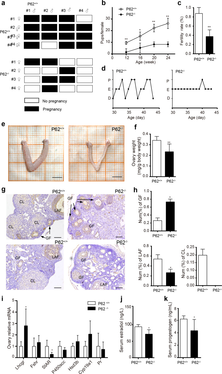
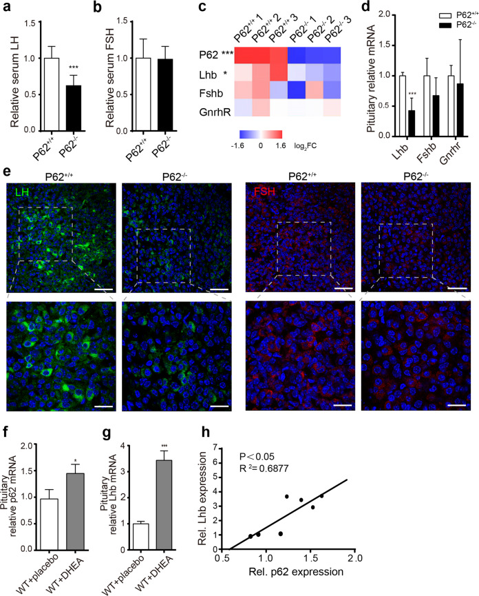
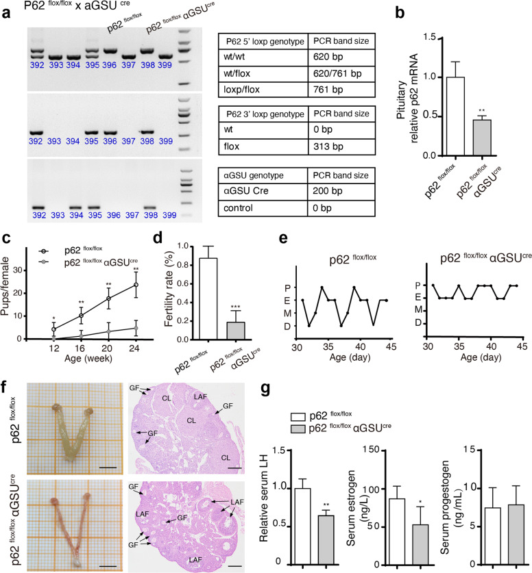
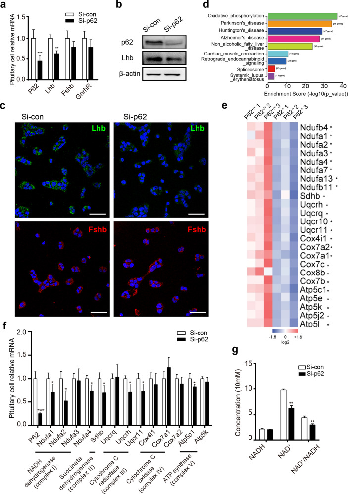
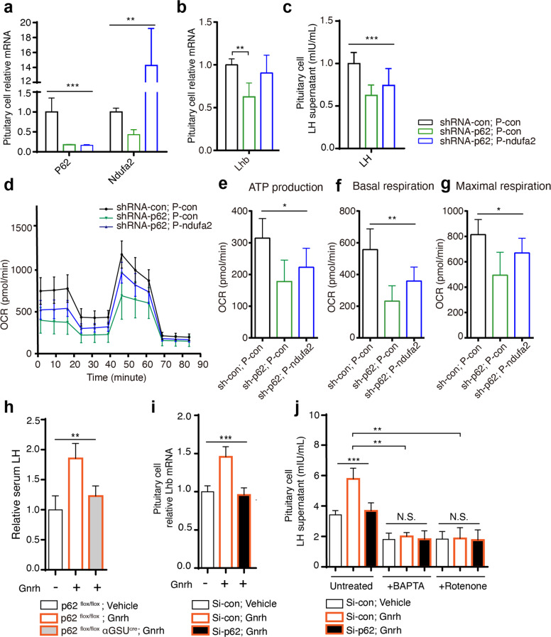
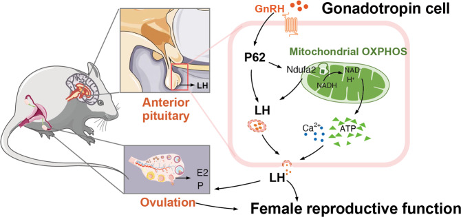
P62 is a protein adaptor for various metabolic processes. Mice that lack p62 develop adult-onset obesity. However, investigations on p62 in reproductive dysfunction are rare. In the present study, we explored the effect of p62 on the reproductive system. P62 deficiency-induced reproductive dysfunction occurred at a young age (8 week old). Young systemic p62 knockout (p62-/-) and pituitary-specific p62 knockout (p62flox/flox αGSUcre) mice both presented a normal metabolic state, whereas they displayed infertility phenotypes (attenuated breeding success rates, impaired folliculogenesis and ovulation, etc.) with decreased luteinizing hormone (LH) expression and production. Consistently, in an infertility model of polycystic ovary syndrome (PCOS), pituitary p62 mRNA was positively correlated with LH levels. Mechanistically, p62-/- pituitary RNA sequencing showed a significant downregulation of the mitochondrial oxidative phosphorylation (OXPHOS) pathway. In vitro experiments using the pituitary gonadotroph cell line LβT2 and siRNA/shRNA/plasmid confirmed that p62 modulated LH synthesis and secretion via mitochondrial OXPHOS function, especially Ndufa2, a component molecule of mitochondrial complex I, as verified by Seahorse and rescue tests. After screening OXPHOS markers, Ndufa2 was found to positively regulate LH production in LβT2 cells. Furthermore, the gonadotropin-releasing hormone (GnRH)-stimulating test in p62flox/flox αGSUcre mice and LβT2 cells illustrated that p62 is a modulator of the GnRH-LH axis, which is dependent on intracellular calcium and ATP. These findings demonstrated that p62 deficiency in the pituitary impaired LH production via mitochondrial OXPHOS signaling and led to female infertility, thus providing the GnRH-p62-OXPHOS(Ndufa2)-Ca2+/ATP-LH pathway in gonadotropic cells as a new theoretical basis for investigating female reproductive dysfunction.


Conflict of interest statement

The authors declare no competing interests.

Figures

Fig. 1
Fig. 1. P62 deficiency leads to female infertility.
a Representative breeding study illustration: four young (8 week old) p62−/− or p62+/+ female mice were paired with four p62+/+ adult male mice for 7 days and then returned to their own cages for 3 weeks to allow for the birth of pups, indicating successful pairing. Each row represents one individual female, and each bar represents one of her pairings. Four examples were chosen from each group to illustrate the fertility phenotypes. b Cumulative number of pups per female and c infertility success rate of female mice in each group during the breeding study, n = 4. d–f Estrous cycles, representative images of ovaries with uteri (scale bar, 500 μm), and the relative quantification of ovary weights in each group. g H&E staining of mouse ovaries. CL corpus luteum, LAF large antral follicle, GF growing follicle; scale bar, 20 μm. h The percentage of ovarian growing follicles, large antral follicles, and corpus luteum counts among all the follicles in each group, n = 4–5. i The relative mRNA expression levels of key steroidogenesis signaling pathway proteins in the ovaries of 8-week-old p62−/− and p62+/+ female mice, n = 4–6. j, k Serum estradiol (E2) and progestogen (P) levels of the young p62−/− and p62+/+ female groups, n = 5–8. The data in i are presented as the mean ± SEM, and the data in the other panels are presented as the mean ± SD. Student’s t-test. *P < 0.05; **P < 0.01.
Fig. 2
Fig. 2. Abnormal p62 expression dysregulated LH production in the pituitary of female mice.
a, b Serum LH and FSH levels of young (8 week old) p62−/− and p62+/+ female mice, n = 6–9. c, d RNA sequencing and RT-PCR detection of the relative expression of gonadotropin markers (Lhb, Fshb, and Gnrhr) in the pituitary. e Representative immunohistological images of pituitary sections stained for LH (green) and FSH (red) of young p62+/+ or p62−/− female mice. DAPI (blue), scale bars: 40 μm (left); 20 μm (right). f, g Relative mRNA expression of p62 and Lhb in the pituitary of PCOS model mice (WT + DHEA) and controls (WT + placebo), n = 3–4. h The correlation between pituitary p62 and relative Lhb mRNA expression in PCOS mice and control mice, n = 7. Statistical analyses were performed using Student’s t-test (a, b, d, f, j) and Pearson analysis (h). Data are presented as the mean ± SD. *P < 0.05; ***P < 0.001.
Fig. 3
Fig. 3. Reproductive dysfunction is exhibited in pituitary-specific p62 knockout mice.
Pituitary-specific p62−/− mice (p62flox/flox αGSUcre) and controls (p62flox/flox) were created using the cre/loxP approach. a Genotyping and PCR bands of p62flox/flox and p62flox/flox αGSUcre mice. b Relative p62 mRNA expression in the pituitary of young female p62flox/flox and p62flox/flox αGSUcre mice, n = 4. c, d Infertility ability was analyzed by counting cumulative pups per female and the successful fertility rate. e The estrous cycle of young female mice, n = 4. f Representative morphology images of the ovaries and uterus by H&E staining of the ovaries in young female p62flox/flox and p62flox/flox αGSUcre mice. GF growing follicle, LAF large antral follicle, CL corpus luteum; Scale bar: 500 μm (left), 200 μm (right). g Serum LH, E2, and P levels in each group, n = 4–6. Statistical analyses were performed using Student’s t-test. Data are shown as the mean ± SD. Student’s t-test. *P ≤ 0.05; **P ≤ 0.01; ***P < 0.001.
Fig. 4
Fig. 4. P62 deficiency leads to downregulated LH and oxidative phosphorylation (OXPHOS) in pituitary gonadotropin cells.
a–c In vitro experiments in which the mouse pituitary gonadotroph LβT2 cell line was transfected with control/p62 siRNA for 48 h. a RT-PCR detection of p62, Lhb, Fshb, and GnRHR mRNA, n = 4–6. b WB detection of silenced p62 expression and LH protein levels. c Immunofluorescence detection of LH (green) and FSH (red) protein expression in LβT2 cells. DAPI in blue, scale bar: 20 μm. d The significant changed pathways in p62−/− mouse pituitaries enriched by KEGG analysis. e Differentially expressed pituitary OXPHOS markers between p62−/− and p62+/+ mice shown as a heatmap from RNA-Seq of young female mice, n = 3. f Relative mRNA expression levels of OXPHOS markers (mitochondrial complex I: Ndufa1–4; complex II: Sdhb; complex III: Uqcrq, Uqcrh, Uqcr11; complex IV: cox4i1, cox7a1, cox7a2; complex V: atp5c1, atp5k) with p62 siRNA treatment, n = 4. g NADH, NAD+ concentration and NAD+ /NADH ratio measurement, n = 4. All data are presented as the mean ± SD. Student’s t-test, *P < 0.05; **P < 0.01; ***P < 0.001.
Fig. 5
Fig. 5. P62 targets the OXPHOS signaling pathway to regulate LH and GnRH-induced LH production.
Rescue experiments were conducted in LβT2 cells by silencing p62 with shRNA, and p62 was stably silenced in cells overexpressing Ndufa2 with a plasmid for 48 h, thus generating three groups of LβT2 cells: shRNA-con, p-con; shRNA-p62, p-con; shRNA-p62, p-Ndufa2. a RT-PCR detection of p62 and Ndufa2 to confirm transfection efficiency, n = 3. b, c mRNA detection of Lhb (n = 3) and measurement of supernatant LH (n = 6–8) in each group. d–g Seahorse assay analysis of mitochondrial respiratory function in LβT2 cells, including ATP production, basal respiration, and maximal respiration, in each group, n = 5. h Relative serum LH in p62flox/flox αGSUcre and p62flox/flox female mice treated with vehicle or the GnRH agonist gonadorelin (7.5 µg/kg body weight), n = 3-4. i Relative Lhb mRNA after gonadorelin induction in LβT2 cells, and j supernatant LH concentration level after treatment with the calcium chelator BAPTA (2 mM) or the mitochondrial NADH: ubiquinone reductase inhibitor rotenone (5 μM), with/without gonadorelin (100 nM), n = 4. All data are presented as the mean ± SD. Statistical analyses were performed using one-way ANOVA and Student’s t-test (b, i). *P < 0.05; **P < 0.01; ***P ≤ 0.001.
Fig. 6
Fig. 6. The modulative role of p62 in female reproductive biology.
Schematic diagram of the GnRH-p62-OXPHOS (Ndufa2)-Ca2+/ATP-LH pathway in pituitary gonadotropic cells for regulating female reproduction.

References

    1. Mascarenhas MN, Flaxman SR, Boerma T, Vanderpoel S, Stevens GA. National, regional, and global trends in infertility prevalence since 1990: a systematic analysis of 277 health surveys. PLoS Med. 2012;9:e1001356. doi: 10.1371/journal.pmed.1001356. - DOI - PMC - PubMed
    1. Mikhael S, Punjala-Patel A, Gavrilova-Jordan L. Hypothalamic-pituitary-ovarian axis disorders impacting female fertility. Biomedicines. 2019;7:1–9. doi: 10.3390/biomedicines7010005. - DOI - PMC - PubMed
    1. World Health Organization. International Classification of Diseases 11th Revision (ICD-11). Executive Board: EB143/13 (WHO, 2018).
    1. Weiss RV, Clapauch R. Female infertility of endocrine origin. Arq. Bras. Endocrinol. Metabol. 2014;58:144–152. doi: 10.1590/0004-2730000003021. - DOI - PubMed
    1. National Collaborating Centre for Women’s and Children’s Health. Fertility: Assessment and Treatment for People with Fertility Problems. NICE Clinical GuidelinesNo. 156 (National Collaborating Centre for Women’s and Children’s Health, 2013).

Publication types

MeSH terms