Review
. 2021 Dec;126(12):1609-1618.
doi: 10.1007/s11547-021-01412-1. Epub 2021 Aug 28.

Phosphaturic mesenchymal tumors: radiological aspects and suggested imaging pathway

Affiliations
Review

Phosphaturic mesenchymal tumors: radiological aspects and suggested imaging pathway

Mohsin A M Hussein et al. Radiol Med. 2021 Dec.

Abstract

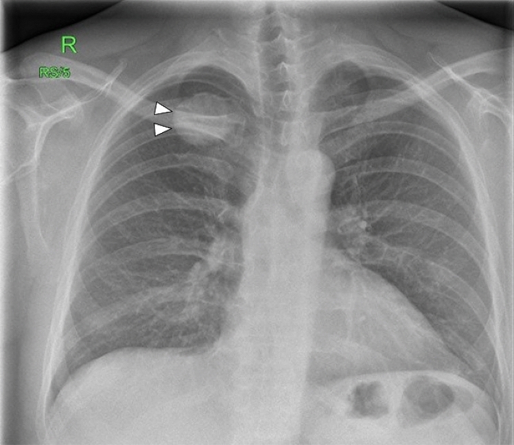
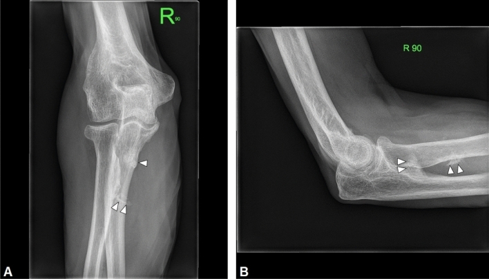
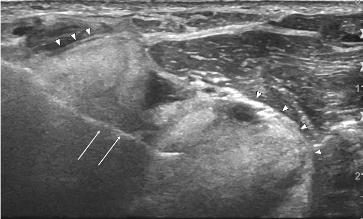
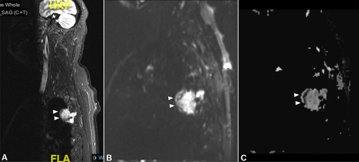
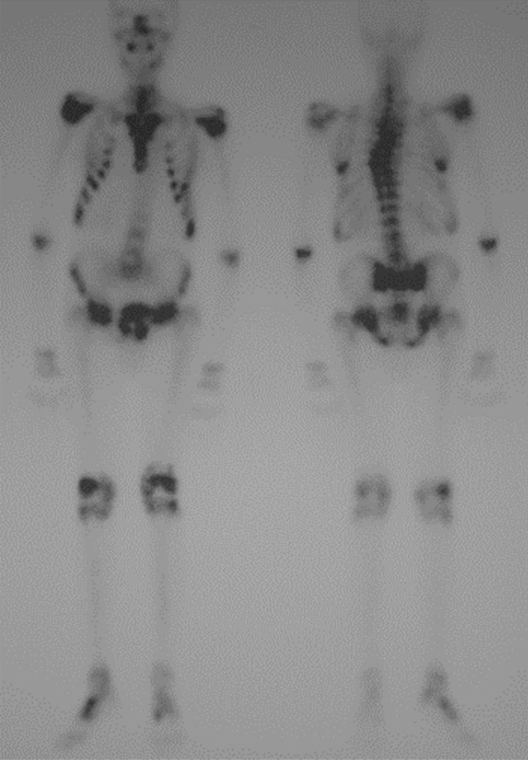
Phosphaturic mesenchymal tumors (PMTs) are rare mesenchymal neoplasms of soft tissue or bone origin that can give rise to a challenge in diagnostic imaging. These tumors are frequently associated with tumor-induced osteomalacia, also called oncogenic osteomalacia, which is a rare paraneoplastic syndrome characterized by ectopic secretion of fibroblast growth factor 23, a hormone that regulates serum phosphate level. PMTs show polymorphic features on both radiological findings and histological examination, causing problems in diagnosis owing to their similarity with other mesenchymal tumors. Thus, this paper aims to describe radiological aspects of PMTs and suggest an imaging pathway for accurate diagnosis throughout the evidence from the literature review.

Keywords: Fibroblast growth factor 23; Oncogenic osteomalacia; Phosphaturic mesenchymal tumors; Tumor-induced osteomalacia.


Conflict of interest statement

The authors declare that they have no conflict of interest.

Figures

Fig. 1
Fig. 1
22-year-old man with a rhinopharyngeal vault PMT who presented with a 4-year history of generalized pain and progressive weakness confining him to a wheelchair. Plain radiograph demonstrates multiple insufficiency fractures (arrowheads) with features of osteomalacia in the feet
Fig. 2
Fig. 2
44-year-old man with a PMT of the foot. Plain radiograph demonstrates a faint increased density in keeping with soft tissue calcification on the plantar aspect of the foot
Fig. 3
Fig. 3
47-year-old man with a PMT of the 4th rib. He presented to the rheumatology service, with an 18-month history of recurrent back pain and unexpected weight loss. On physical examination, he had reduced spinal movement and point tenderness over his sacroiliac joints. Chest X-ray shows a well-defined rounded dense lesion in the 4th rib, with uniform calcification
Fig. 4
Fig. 4
65-year-old man with a PMT of the right elbow. AP (A) and lateral (B) radiographs of the shoulder demonstrate an ill-defined area of soft tissue calcification in the proximal forearm
Fig. 5
Fig. 5
65-year-old man with a PMT of the right elbow. Ultrasound of the proximal forearm shows a large extraosseous component (arrow heads) which was not apparent on plain radiograph. Biopsy needle (arrows)
Fig. 6
Fig. 6
22-year-old man with a rhinopharyngeal vault PMT who presented with a 4-year history of generalized pain and progressive weakness confining him to a wheelchair. (A) Axial CT shows an enhancing bulging tumor arising from the left ethmoidal sinus with an extraosseous soft tissue component. Axial (B) and coronal (C) axial T2 weight spin-echo MR image show a lesion of the left ethmoidal sinus demonstrating an increased signal intensity of the solid mass, with multiple dark foci or vascular flow voids
Fig. 7
Fig. 7
47-year-old man with a PMT of the 4th rib. (A, B) Axial CT demonstrates a well-defined rounded dense lesion with a mildly calcified internal matrix. (C) Axial T2 weighted spin-echo MR image shows a lesion of the 4th rib with mixed hyper- and hypointense signal, well-defined borders, cortical destruction, and extension into the extrapleural fat. (D) Axial T1 weighted fat suppressed contrast enhanced image shows intense uniform enhancement of the lesion. The non-enhancing margins (arrowheads) are areas of calcification
Fig. 8
Fig. 8
47-year-old man with a PMT of the 4th rib. Para-sagittal STIR sequence (A) demonstrates high signal intensity mass with an anterior rim of low signal intensity, arising from the rib. Intense intramedullary edema is noted of the rib. Diffusion-weighted imaging (B) with an apparent diffusion coefficient map (C) demonstrates restricted diffusion along the anterior margin in keeping with the calcified rim of the PMT
Fig. 9
Fig. 9
22-year-old man with a rhinopharyngeal vault PMT. Whole-body bone scintigraphy demonstrated multiple areas of increased uptake in the thoracic spine, ribs, pelvis, and limbs with a characteristic H-shape pattern in the sacrum, typically found in insufficiency fractures
Fig. 10
Fig. 10
Imaging pathway for PMTs

References

    1. Kaul M, Silverberg M, Dicarlo EF, Schneider R, Bass AR, Erkan D. Tumor-induced osteomalacia. Clin Rheumatol. 2007;26(9):1575–1579. - PubMed
    1. Shimada T, et al. FGF-23 is a potent regulator of vitamin D metabolism and phosphate homeostasis. J Bone Miner Res. 2004;19(3):429–435. - PubMed
    1. Florenzano P, Hartley IR, Jimenez M, Roszko K, Gafni RI, Collins MT. Tumor-induced osteomalacia. Calcif Tissue Int. 2021;108(1):128–142. - PubMed
    1. McCANCE RA. Osteomalacia with Looser's nodes (Milkman's syndrome) due to a raised resistance to vitamin D acquired about the age of 15 years. Q J Med. 1947;16(1):33–46. - PubMed
    1. Shevroja E, Cafarelli FP, Guglielmi G, Hans D. DXA parameters, trabecular bone score (TBS) and bone mineral density (BMD), in fracture risk prediction in endocrine-mediated secondary osteoporosis. Endocrine. 2021 doi: 10.1007/s12020-021-02806-x. - DOI - PMC - PubMed

MeSH terms

Supplementary concepts