. 2021 Oct;25(19):9411-9426.
doi: 10.1111/jcmm.16883. Epub 2021 Aug 28.

KCTD11 inhibits progression of lung cancer by binding to β-catenin to regulate the activity of the Wnt and Hippo pathways

Affiliations

KCTD11 inhibits progression of lung cancer by binding to β-catenin to regulate the activity of the Wnt and Hippo pathways

Man Yang et al. J Cell Mol Med. 2021 Oct.

Abstract

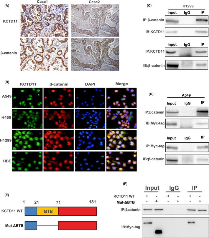
KCTD11 has been reported to be a potential tumour suppressor in several tumour types. However, the expression of KCTD11 and its role has not been reported in human non-small cell lung cancer (NSCLC). Whether its potential molecular mechanism is related to its BTB domain is also unknown. The expression of KCTD11 in 139 NSCLC tissue samples was detected by immunohistochemistry, and its correlation with clinicopathological factors was analysed. The effect of KCTD11 on the biological behaviour of lung cancer cells was verified in vitro and in vivo. Its effect on the epithelial-mesenchymal transition(EMT)process and the Wnt/β-catenin and Hippo/YAP pathways were observed by Western blot, dual-luciferase assay, RT-qPCR, immunofluorescence and immunoprecipitation. KCTD11 is under-expressed in lung cancer tissues and cells and was negatively correlated with the degree of differentiation, tumour-node-metastasis (TNM) stage and lymph node metastasis. Low KCTD11 expression was associated with poor prognosis. KCTD11 overexpression inhibited the proliferation and migration of lung cancer cells. Further studies indicated that KCTD11 inhibited the Wnt pathway, activated the Hippo pathway and inhibited EMT processes by inhibiting the nuclear translocation of β-catenin and YAP. KCTD11 lost its stimulatory effect on the Hippo pathway after knock down of β-catenin. These findings confirm that KCTD11 inhibits β-catenin and YAP nuclear translocation as well as the malignant phenotype of lung cancer cells by interacting with β-catenin. This provides an important experimental basis for the interaction between KCTD11, β-catenin and YAP, further revealing the link between the Wnt and Hippo pathways.

Keywords: Hippo pathway; KCTD11; Wnt pathway; YAP; β-catenin.


Conflict of interest statement

The authors declare that they have no competing interests.

Figures

FIGURE 1
FIGURE 1
Low expression of KCTD11 in lung cancer is associated with poor prognosis. (A, B) The expression levels of KCTD11 are downregulated in 20 cases of NSCLC tissues compared to normal tissues, as assessed by Western blot (A) and analysed with GraphPad prism (B). (C) Immunohistochemical staining of KCTD11 in the surrounding normal tissues and the representative carcinoma (n = 139). KCTD11 is expressed in the nuclei and cytoplasm and show strong positive expression in normal alveolar cells (a) and bronchial epithelial cells (b); positive expression of KCTD11 in highly differentiated adenocarcinoma (c) and squamous cell carcinoma (e); negative expression of KCTD11 in poorly differentiated adenocarcinoma (d) and squamous cell carcinoma (f) is observed (Magnification:×200). (D) Western blot is used to detect the protein level of KCTD11 in eight lung cell lines. (E) The data from Kaplan‐Meier database show that the survival time of KCTD11‐positive patients is significantly longer than that of KCTD11‐negative patients (p  <  0.05). GAPDH and β‐Actin serve as the loading control
FIGURE 2
FIGURE 2
KCTD11 inhibits proliferation and invasion of NSCLC. (A–C) In KCTD11‐overexpressing A549 cells, a colony formation assay, Transwell assay and MTT assay show that the proliferation and invasion of cells are reduced, (D–F) whereas the proliferation and invasion of cells are increased in KCTD11‐depleted H1299 cells. (G‐I) KCTD11 overexpression inhibits tumour formation and metastases in nude mice. Subcutaneous injection of KCTD11‐overexpressing A549 cells (G418 screening) into nude mice (n = 3) and the control group (n = 3). The mice are sacrificed, and an autopsy performed to examine the growth and spread of the tumour after six weeks. Compared with the control group, the tumour formation is reduced. (J) Mice injected with KCTD11‐overexpressing A549 cells through tail vein have decreased pulmonary metastases than those in the control group, *p  <  0.05. (K‐L) The transfection efficiency is shown by Western blot after overexpressing or interfering KCTD11. All the experiments are repeated three times independently, and the results are the mean. Abbreviations: EV, Empty vector; NC, Negative control
FIGURE 3
FIGURE 3
KCTD11 activates the Hippo pathway by upregulating the phosphorylation level of YAP in NSCLC cells. (A, B) KCTD11 overexpression inhibits the transcriptional activity of TEAD. By Hippo pGTII luciferase reporter, transfection of KCTD11 in A549 cells upregulates the activity of Hippo signalling pathway, and depleted KCTD11 in H1299 cells inhibits the activity of Hippo signalling pathway. The experiment is treated with control or wnt3a conditioned medium for 6 h. (C, D) The target mRNA expression levels of the Hippo pathway, CTGF, CYR61 and CyclinE are downregulated in KCTD11‐overexpressing A549 cells and are upregulated in KCTD11‐depleted H1299 cells. (E, F) Western blot shows that the phosphorylation of YAP is increased, and expression levels of target genes of the Hippo pathway, CTGF and CyclinE are downregulated in KCTD11‐overexpressing A549 cells and H460 cells. In KCTD11‐depleted H1299 cells and HBE cells, the phosphorylation of YAP decreases, and expression levels of CTGF and CyclinE are upregulated. GAPDH is used as a loading control. The grey value is analysed by using Image J software. (G–I) Nucleus‐cytoplasm isolation and immunofluorescence demonstrate that the nuclear translocation of YAP is inhibited in A549 cell lines after transfection of KCTD11 and silencing of KCTD11 in H1299 cells leads to an increase in the nuclear translocation of YAP. (I, J, magnification 400×; Bars: 20 µm). Results are shown from three independent experiments
FIGURE 4
FIGURE 4
KCTD11 inhibits the Wnt pathway and nuclear translocation of β‐catenin. (A, B) By Wnt TOPflash reporter, transfection of KCTD11 in A549 cells downregulates the activity of Wnt signalling pathway, and depleted KCTD11 in H1299 cells upregulates the activity of Wnt signalling pathway. The experiment is treated with control or wnt3a conditioned medium for 6 h. (C, D) The target genes mRNA expression levels of the Wnt pathway, MMP7, CyclinD1 and Axin2 are downregulated in KCTD11‐overexpressing A549 cells and are upregulated in KCTD11‐depleted H1299 cells. (E, F) Western blot shows that the levels of β‐catenin and the transcriptional activity of Wnt pathway downstream target genes, C‐myc, cyclin D1 and MMP7, are downregulated in KCTD11‐overexpressing A549 cells and H460 cells but upregulated in KCTD11‐depleted H1299 cells and HBE cell. GAPDH is used as a loading control. The grey value is analysed by using Image J software. (G–I) Nucleus‐cytoplasm isolation and immunofluorescence demonstrate that the nuclear translocation of β‐catenin is decreased in A549 cell lines after transfection of KCTD11 and silencing of KCTD11 in H1299 cells leads to an addition in the nuclear translocation of β‐catenin. (I, J, magnification 400×; Bars: 20 µm). Results are shown from three independent experiments
FIGURE 5
FIGURE 5
KCTD11 inhibits the process of EMT. (A, B) A549 cells and H460 cells are transfected with KCTD11, the cells are lysed 48 h later, and the level of proteins are detected by Western blot. E‐cadherin and ZO‐1 are upregulated while N‐cadherin, vimentin, Snail and Slug are downregulated in KCTD11‐overexpressing A549 cells and H460 cells, while the results are reversed in KCTD11‐depleted H1299 cells and HBE cells. GAPDH is used as a loading control. (C, D) KCTD11 induces a morphology change and representative phase‐contrast images of cells growing in monolayer cultures. KCTD11 transfection preserves the cobblestone‐like appearance of tight intercellular junctions in A549 cells, while depleting endogenous KCTD11 in H1299 cells show the opposite effect, with the cells exhibiting a spindle‐like morphology (magnification, 200×;Bars: 50 µm)
FIGURE 6
FIGURE 6
KCTD11 binds to β‐catenin via the BTB domain. (A) Immunohistochemical staining of serial sections and statistical analysis to show that KCTD11 is negatively correlated with β‐catenin expression (p = 0.035). (B) Immunofluorescence reveals that endogenous KCTD11 and β‐catenin had co‐localization in NSCLC cell lines. (C) Interaction between endogenous KCTD11 and β‐catenin is detected by co‐immunoprecipitation in A549 cells. Cell lysates are immunoprecipitated with anti‐KCTD11 antibody or anti‐β‐catenin, and then, the expression level of KCTD11 and β‐catenin by immunoblotting is examined. The IgG serves as negative control. (D) The interaction between exogenous KCTD11 and β‐catenin is also detected by immunoprecipitation with transfection of Myc‐tagged KCTD11 plasmids into A549 cells. (E) Different locations within the structure of wild KCTD11 plasmid and Mutant MYC‐KCTD11‐ΔBTB (mut‐ΔBTB) plasmid. (F) KCTD11interacts with β‐catenin via its BTB domain. A549 cells are transfected with KCTD11 and mut‐ΔBTB plasmid, and the resultant cell lysates are immunoprecipitated with anti‐β‐catenin antibody. Deletion of BTB domain preventing KCTD11 from binding to β‐catenin is confirmed by the absence of Myc‐tag. (magnification, 200×;Bars: 50 µm)
FIGURE 7
FIGURE 7
KCTD11 regulates Hippo pathway activity by β‐catenin. (A) Transfection of KCTD11 in A549 and H460 cell lines, Western blot showing that the expression level of active β‐catenin is downregulated and P‐β‐catenin is upregulated. (B) The expression level of active β‐catenin is upregulated, and P‐β‐catenin is downregulated after silencing of KCTD11 in H1299 and HBE cells. (C) KCTD11 inhibits YAP through β‐catenin. A549 cells are transfected with KCTD11 and EV, and si‐catenin and siNC. Western blot showing that after knocking out β‐catenin, the expression levels of YAP and P‐YAP are reduced and that overexpression of KCTD11 could not reverse this change, with the level of YAP and P‐YAP still being inhibited. (D, E) KCTD11 acts as a tumour suppressor through the BTB domain. A549 cells are transfected with EV, KCTD11 and mut‐ΔBTB plasmid and the level of proteins are detected by Western blot. Overexpression of mut‐ΔBTB does not affect the expression of Hippo and wnt pathway‐related proteins and EMT‐related proteins. (F) The Wnt TOPflash reporter and Hippo pGTII luciferase reporter are used to detect the transcriptional activity of YAP / TEAD and β‐catenin / TCF‐4 after transfection of mut‐ΔBTB. Abbreviations: EV, empty vector; siNC, negative control siRNAs

Similar articles

Cited by

References

    1. Poon CLC, Liu W, Song Y, et al. A Hippo‐like signaling pathway controls tracheal morphogenesis in drosophila melanogaster. Dev Cell. 2018;47(5):564‐575.e5. 10.1016/j.devcel.2018.09.024 - DOI - PMC - PubMed
    1. Yu FX, Zhao B, Guan KL. Hippo pathway in organ size control, tissue homeostasis, and cancer. Cell. 2015;163(4):811‐828. 10.1016/j.cell.2015.10.044 - DOI - PMC - PubMed
    1. Lin KC, Moroishi T, Meng Z, et al. Regulation of Hippo pathway transcription factor TEAD by p38 MAPK‐induced cytoplasmic translocation. Nat Cell Biol. 2017;19(8):996‐1002. 10.1038/ncb3581 - DOI - PMC - PubMed
    1. Janse van Rensburg HJ, Azad T, Ling M, et al. The Hippo pathway component TAZ promotes immune evasion in human cancer through PD‐L1. Cancer Res. 2018;78(6):1457‐1470. 10.1158/0008-5472.can-17-3139 - DOI - PubMed
    1. Marti P, Stein C, Blumer T, et al. YAP promotes proliferation, chemoresistance, and angiogenesis in human cholangiocarcinoma through TEAD transcription factors. Hepatology. 2015;62(5):1497‐1510. 10.1002/hep.27992 - DOI - PubMed

Publication types

MeSH terms