. 2021 Sep;20(9):e13457.
doi: 10.1111/acel.13457. Epub 2021 Aug 28.

Genetic reduction of mTOR extends lifespan in a mouse model of Hutchinson-Gilford Progeria syndrome

Affiliations

Genetic reduction of mTOR extends lifespan in a mouse model of Hutchinson-Gilford Progeria syndrome

Wayne A Cabral et al. Aging Cell. 2021 Sep.

Abstract

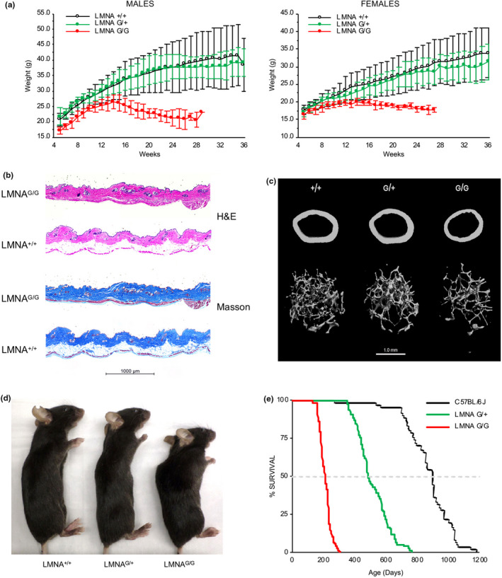
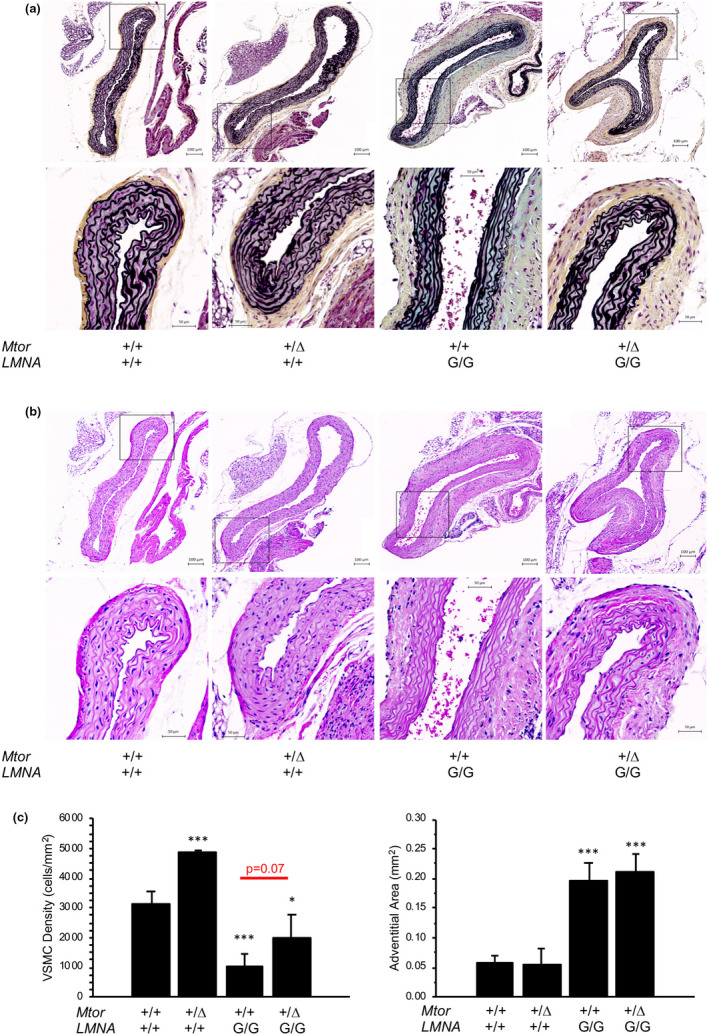
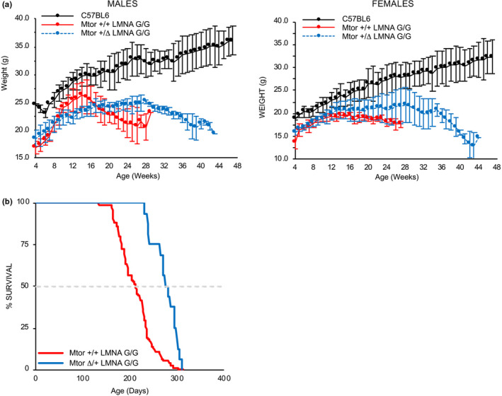
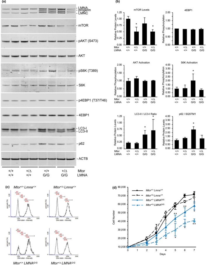
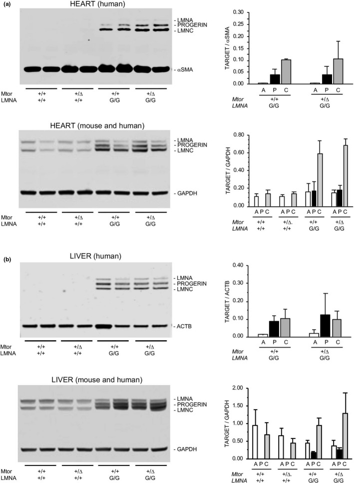
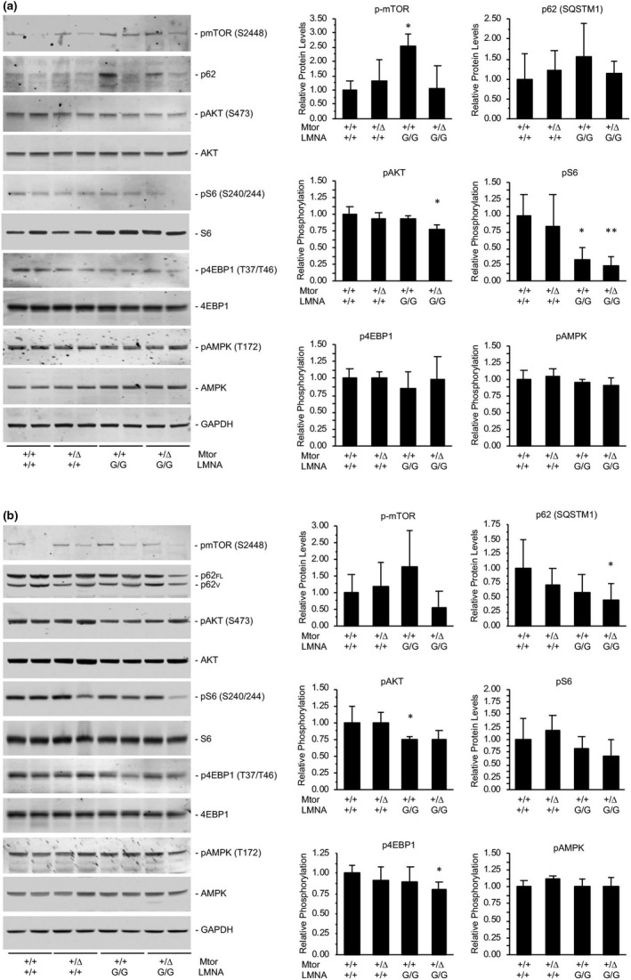
Hutchinson-Gilford progeria syndrome (HGPS) is a rare accelerated aging disorder most notably characterized by cardiovascular disease and premature death from myocardial infarction or stroke. The majority of cases are caused by a de novo single nucleotide mutation in the LMNA gene that activates a cryptic splice donor site, resulting in production of a toxic form of lamin A with a 50 amino acid internal deletion, termed progerin. We previously reported the generation of a transgenic murine model of progeria carrying a human BAC harboring the common mutation, G608G, which in the single-copy state develops features of HGPS that are limited to the vascular system. Here, we report the phenotype of mice bred to carry two copies of the BAC, which more completely recapitulate the phenotypic features of HGPS in skin, adipose, skeletal, and vascular tissues. We further show that genetic reduction of the mechanistic target of rapamycin (mTOR) significantly extends lifespan in these mice, providing a rationale for pharmacologic inhibition of the mTOR pathway in the treatment of HGPS.

Keywords: S6 Kinase; lamin A/C; laminopathies; mTOR; progeria.


Conflict of interest statement

The authors declare no competing interests.

Figures

FIGURE 1
FIGURE 1
Murine model harboring two copies of the human LMNA G608G transgene develops a severe progeroid phenotype. (a) Growth curves of wild‐type (LMNA +/+; n = 16 males, 13 females), mice carrying one copy (LMNA G/+; n = 8 males, 8 females), or two copies (LMNA G/G; n = 9 males, 8 females) of the human transgene containing the LMNA G608G mutation. By 5 weeks of age, LMNA G/G weigh significantly less than LMNA +/+ and LMNA G/+ littermates. Males, 5–15 weeks < 0.01, 16–28 weeks < 0.001; Females, 5–15 weeks < 0.05, 16–23 weeks < 0.01, 24–27 weeks < 0.001. (b) Histologic analysis of skin from LMNA G/G and wild‐type (LMNA +/+) littermates at 6 months of age demonstrates decreased subcutaneous fat in transgenic mice. Skin sections were stained with hematoxylin and eosin (H&E) and Masson's trichrome, which reveals the keratin and muscle (red), collagen (blue), and cellular cytoplasm (pink) and nuclei (dark brown). The unstained adipose tissue is located between the collagenous dermal layers (blue) and dark red‐stained panniculus carnosus (fast twitch type IIB glycolytic fibers). (c) Micro‐computed tomography (μCT)‐derived images of femoral bone of wild‐type (+/+), single‐copy (G/+) and double‐copy (G/G) transgenic mice at 6 months of age. Both cortical (top images) and trabecular (bottom images) structural parameters demonstrate the reduced bone volume in LMNA G/G mice. (d) Lateral images of 5‐month‐old mice reveal kyphosis and growth deficiency observed in double‐copy (LMNA G/G) transgenic mice compared to wild‐type (LMNA +/+) and single‐copy (LMNA G/+) mice. (e) Kaplan‐Meier plots illustrating the shortened lifespan of single‐copy (LMNA G/+, n = 79) and double‐copy (LMNA G/G, n = 181) transgenic mice relative to wild type (LMNA +/+; C57BL/6J, n = 61)
FIGURE 2
FIGURE 2
Severe vascular phenotype in HGPS mice can be partially rescued by genetic reduction of Mtor. (a) Movat's pentachrome‐stained ascending aorta sections from 5‐month‐old mice. (b) Hematoxylin and eosin (H&E)‐stained aortae from LMNA +/+ and LMNA G/G mice. (c) Quantitation of adventitial area and VSMC number in aortae. Genetic reduction of Mtor partially restores the loss of vascular smooth muscle cells but does not rescue the adventitial expansion of aortae in LMNA G/G mice. *, < 0.05; **, < 0.01; ***, < 0.001
FIGURE 3
FIGURE 3
Genetic reduction of Mtor extends lifespan in LMNA G608G transgenic mice. (a) Growth curves of wild‐type (Mtor +/+ LMNA +/+), and double‐copy transgenic mice containing two wild‐type mTor alleles (Mtor +/+ LMNA G/G) or heterozygous for the Mtor hypomorphic allele (Mtor Δ /+ LMNA G/G). Growth deficiency in LMNA G/G mice is not rescued by reduced expression of mTOR in male (n = 6 Mtor +/+ LMNA +/+, 7 Mtor +/+ LMNA G/G, 13 Mtor Δ /+ LMNA G/G) or female (n = 6 Mtor +/+ LMNA +/+, 6 Mtor +/+ LMNA G/G, 12 Mtor Δ /+ LMNA G/G) mice. (b) Kaplan‐Meier plot demonstrates the 30% extension in lifespan of double‐copy transgenic mice heterozygous for the Mtor hypomorphic allele (Mtor Δ /+ LMNA G/G) compared to transgenic mice harboring two wild‐type Mtor alleles (Mtor +/+ LMNA G/G). p < 0.001
FIGURE 4
FIGURE 4
Reduction of mTOR levels in LMNA G608G newborn fibroblasts normalizes S6K activity. (a) Western analyses of A‐type lamins and mTOR signaling pathway components in fibroblast cell lines derived from Mtor +/+ LMNA +/+, Mtor Δ /+ LMNA +/+, Mtor +/+ LMNA G/G, and Mtor Δ /+ LMNA G/G mice. (b) Quantitative analysis of mTOR signaling components from immunoblots demonstrates reduction of mTOR protein in cells heterozygous for the Mtor hypomorphic allele. Induction of autophagy is indicated by increased LC3‐II/I ratios in Mtor +/+ LMNA G/G and Mtor Δ /+ LMNA G/G cells versus wild‐type, and reduced levels of p62 in Mtor Δ /+ LMNA G/G cells versus Mtor +/+ LMNA G/G cells. Normalization of increased S6K phosphorylation in LMNA G/G cells occurs with reduced mTOR levels. *, < 0.05 versus Mtor +/+ LMNA +/+ (c) Cell cycle analysis of proliferative fibroblasts in culture. M1, G0/G1 phase; M2, S phase; M3, G2/M phase. Genetic reduction of Mtor in transgenic cells (Mtor Δ /+ LMNA G/G) shifts the fraction of cells in G0/G1, S, and G2/M phases to levels seen in wild‐type (Mtor +/+ LMNA +/+) cells. (d) LMNA G/G fibroblasts are less proliferative than LMNA +/+ cells in culture. Heterozygosity for the Mtor hypomorphic allele further inhibits proliferation of LMNA G/G murine fibroblasts. *, < 0.05; **, < 0.01; Mtor +/+ LMNA G/G significance versus Mtor +/+ LMNA +/+; Mtor Δ /+ LMNA G/G significance versus Mtor +/+ LMNA G/G
FIGURE 5
FIGURE 5
Analysis of A‐type lamins in tissues. (a, b) Western analysis of A‐type lamins extracted from heart tissue and livers of Mtor +/+ LMNA +/+, Mtor Δ /+ LMNA +/+, Mtor +/+ LMNA G/G, and Mtor Δ /+ LMNA G/G mice (n = 2 per genotype, 1 male and 1 female). Samples in upper immunoblots were immunoprecipitated with antibodies to human lamin A/C [JoL2] and smooth muscle actin (SMA) or beta‐actin (ACTB), then probed with an alternative antibody recognizing both mouse and human Lamin A/C and reference proteins SMA or ACTB. Lower immunoblots contain tissue homogenates probed with antibody recognizing both mouse and human Lamin A/C [4c11] and GAPDH. No significant difference in lamin A, progerin, or lamin C levels was observed in mice with genetic reduction of mTOR regardless of sample preparation procedure, antibody used for detection, or reference protein used for normalization
FIGURE 6
FIGURE 6
mTOR signaling pathway is inhibited in adult LMNA G608G mice in vivo. (a) Representative immunoblots of mTOR signaling pathway components isolated from heart tissue of 5 month‐old Mtor +/+ LMNA +/+, Mtor Δ /+ LMNA +/+, Mtor +/+ LMNA G/G, and Mtor Δ /+ LMNA G/G mice. Despite hyperactivation of mTOR in Mtor +/+ LMNA G/G, relative phosphorylation of S6 decreased versus wild type. (b) Western analyses of liver homogenates. Genetic reduction of Mtor significantly reduces levels of p62 isoforms (p62FL, full length; p62V, variant (Kageyama et al., 2018)) and phosphorylated 4EBP1 in LMNA G/G mice. N = 4 mice per genotype (2 males, 2 females); *, < 0.05; **, < 0.01, Mtor +/+ LMNA G/G and Mtor Δ /+ LMNA G/G significance versus Mtor +/+ LMNA +/+

References

    1. Cao, K., Graziotto, J. J., Blair, C. D., Mazzulli, J. R., Erdos, M. R., Krainc, D., & Collins, F. S. (2011). Rapamycin reverses cellular phenotypes and enhances mutant protein clearance in Hutchinson‐Gilford progeria syndrome cells. Science Translational Medicine, 3(89), 89ra58. 10.1126/scitranslmed.3002346 - DOI - PubMed
    1. Capell, B. C., Olive, M., Erdos, M. R., Cao, K., Faddah, D. A., Tavarez, U. L., Conneely, K. N., Qu, X., San, H., Ganesh, S. K., Chen, X., Avallone, H., Kolodgie, F. D., Virmani, R., Nabel, E. G., & Collins, F. S. (2008). A farnesyltransferase inhibitor prevents both the onset and late progression of cardiovascular disease in a progeria mouse model. Proceedings of the National Academy of Sciences, 105(41), 15902–15907. 10.1073/pnas.0807840105 - DOI - PMC - PubMed
    1. Cenni, V., Capanni, C., Columbaro, M., Ortolani, M., D'Apice, M. R., Novelli, G., Fini, M., Marmiroli, S., Scarano, E., Maraldi, N. M., Squarzoni, S., Prencipe, S., & Lattanzi, G. (2011). Autophagic degradation of farnesylated prelamin A as a therapeutic approach to lamin‐linked progeria. European Journal of Histochemistry, 55(4), e36. 10.4081/ejh.2011.e36 - DOI - PMC - PubMed
    1. Cheng, K. W., Agarwal, R., Mitra, S., Lee, J. S., Carey, M., Gray, J. W., & Mills, G. B. (2012). Rab25 increases cellular ATP and glycogen stores protecting cancer cells from bioenergetic stress. EMBO Molecular Medicine, 4(2), 125–141. 10.1002/emmm.201100193 - DOI - PMC - PubMed
    1. Cubria, M. B., Suarez, S., Masoudi, A., Oftadeh, R., Kamalapathy, P., DuBose, A., Erdos, M. R., Cabral, W. A., Karim, L., Collins, F. S., Snyder, B. D., & Nazarian, A. (2020). Evaluation of musculoskeletal phenotype of the G608G progeria mouse model with lonafarnib, pravastatin, and zoledronic acid as treatment groups. Proceedings of the National Academy of Sciences, 117(22), 12029–12040. 10.1073/pnas.1906713117 - DOI - PMC - PubMed

Publication types

Substances

LinkOut - more resources