. 2021 Nov;142(5):807-825.
doi: 10.1007/s00401-021-02361-9. Epub 2021 Aug 28.

Apolipoprotein E regulates lipid metabolism and α-synuclein pathology in human iPSC-derived cerebral organoids

Affiliations

Apolipoprotein E regulates lipid metabolism and α-synuclein pathology in human iPSC-derived cerebral organoids

Jing Zhao et al. Acta Neuropathol. 2021 Nov.

Erratum in

Abstract

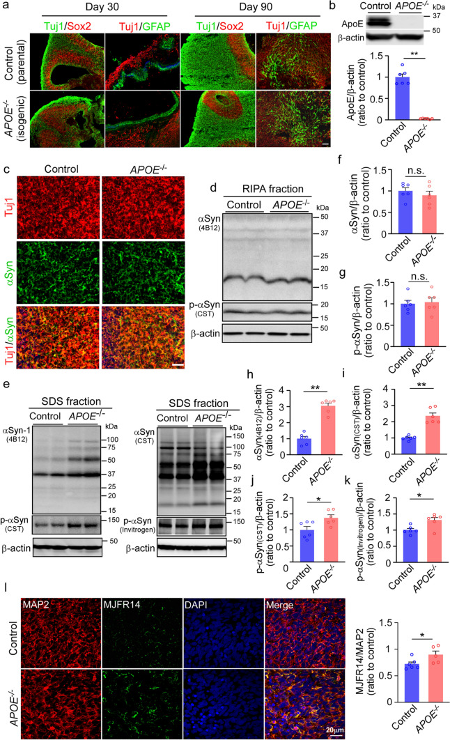
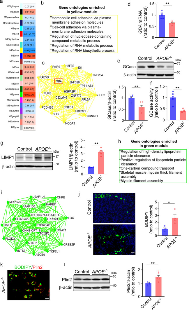
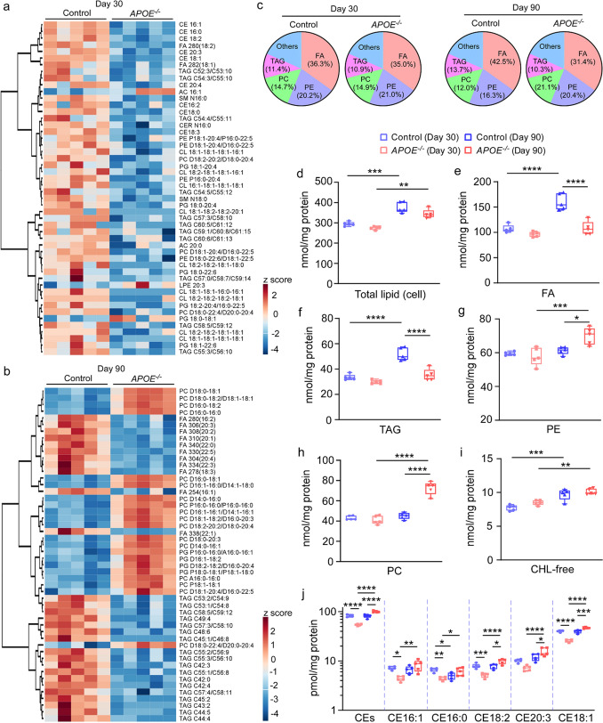
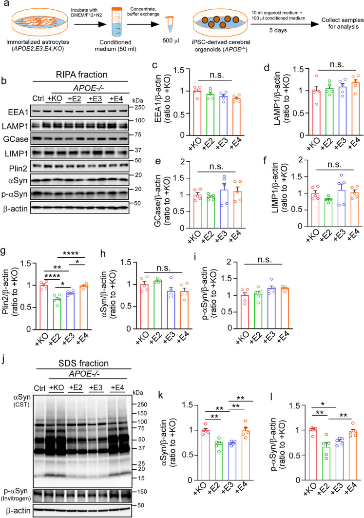
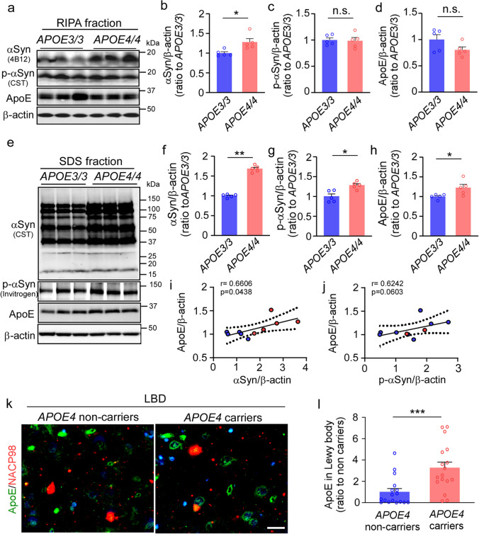
APOE4 is a strong genetic risk factor for Alzheimer's disease and Dementia with Lewy bodies; however, how its expression impacts pathogenic pathways in a human-relevant system is not clear. Here using human iPSC-derived cerebral organoid models, we find that APOE deletion increases α-synuclein (αSyn) accumulation accompanied with synaptic loss, reduction of GBA levels, lipid droplet accumulation and dysregulation of intracellular organelles. These phenotypes are partially rescued by exogenous apoE2 and apoE3, but not apoE4. Lipidomics analysis detects the increased fatty acid utilization and cholesterol ester accumulation in apoE-deficient cerebral organoids. Furthermore, APOE4 cerebral organoids have increased αSyn accumulation compared to those with APOE3. Carrying APOE4 also increases apoE association with Lewy bodies in postmortem brains from patients with Lewy body disease. Our findings reveal the predominant role of apoE in lipid metabolism and αSyn pathology in iPSC-derived cerebral organoids, providing mechanistic insights into how APOE4 drives the risk for synucleinopathies.


Conflict of interest statement

G.B. consults for SciNeuro and E-Scape, and had consulted for AbbVie and Eisai. All other authors declare no competing interests.

Figures

Fig. 1
Fig. 1
Exacerbated α-synuclein accumulation in apoE-deficient cerebral organoids. Parent control and isogenic APOE knockout (APOE−/−) iPSCs were differentiated into cerebral organoids. a, Representative images of the ventricular zone (VZ)-like structure (Tuj1, neuronal marker; SOX2, neural progenitor cell marker; and GFAP, astrocyte maker) in cerebral organoids at Day 30 and Day 90 of differentiation. Scale bar: 100 μm. b, ApoE depletion in isogenic APOE−/− iPSC-derived cerebral organoids were confirmed by Western blotting at Day 90. c, Representative images of α-synuclein immunoreactivity in neurons of iPSC-derived cerebral organoids. Scale bar: 100 μm. d–g, Amounts of total α-synuclein (αSyn; f) and phosphorylated αSyn (p-αSyn; g) in the RIPA soluble fractions of cerebral organoids were quantified by Western blotting (d). ek, Amounts of total αSyn (eh, i) and p-αSyn (ej, k) in the SDS-soluble fractions of cerebral organoids were quantified by Western blotting using two sets of different antibodies. ApoE, αSyn and p-αSyn levels were normalized to β-actin levels. 3 cerebral organoids were pooled and analyzed as one sample. All data are expressed as mean ± SEM (n = 6 samples/each). Experiments were repeated in three independent differentiation batches. l, Amounts of aggregated αSyn in cerebral organoids were quantified by MJFR14 (αSyn aggregate antibody) immunostaining. Scale bar: 20 μm. All data are expressed as mean ± SEM (n = 5–6 organoids/each). Mann–Whitney U tests were performed to determine statistical significance. *p < 0.05, **p < 0.01, n.s., not significant
Fig. 2
Fig. 2
Altered transcriptional profiles of apoE-deficient cerebral organoids implicating GBA and lipid-related pathways. RNA-seq was performed on parental control and isogenic APOE−/− iPSC-derived cerebral organoids at Day 90 (3 cerebral organoids were pooled and analyzed as one sample, n = 3 samples/each). a, Module-trait relationships between groups revealed by WGCNA are shown. The correlation coefficient (r) and the correlation p-value in the parentheses are indicated in each module. Orange indicates upregulation in APOE−/− organoids; blue indicates downregulation in APOE−/− organoids (upregulation in controls) b, Top gene ontologies enriched by the yellow module genes. c, Interaction of top 20 genes with the highest connectivity among each other in the yellow module. d–e, GBA mRNA expression (d) and β-glucocerebrosidase (GCase) levels in the RIPA lysates (e) were quantified by RT-qPCR and Western blotting at Day 90, respectively. f, GCase activity in cerebral organoids was detected by GCase activity kit (Fluorometric). Data were normalized to protein concentrations. g, Amounts of LIMP1 in the iPSC-derived cerebral organoids at Day 90 were quantified by Western blotting. h, Top gene ontologies enriched by the green module genes. (i) Interaction of top 20 genes with the highest connectivity among each other in the green module. j, The lipid droplet accumulation was evaluated by BODIPY staining (Scale bar: 20 μm) with the fluorescent intensity quantified by Image J. All data are expressed as mean ± SEM (n = 4 organoids/each). k, Representative images of co-staining of BODIPY and Plin2 (lipid droplet membrane marker). Scale bar: 20 μm. l, Amounts of Plin2 in the iPSC-derived cerebral organoids at Day 90 were quantified by Western blotting. 3 cerebral organoids were pooled and analyzed as one sample. All data are expressed as mean ± SEM (n = 6 samples/each). Experiments were repeated in three independent differentiation batches. Mann–Whitney U tests were performed to determine statistical significance. *p < 0.05, **p < 0.01
Fig. 3
Fig. 3
Impaired lipid profiles in apoE-deficient cerebral organoids. Lipidomics and cholesterol assay were performed on lysates from parental control and isogenic APOE−/− iPSC-derived cerebral organoids at Day 30 and Day 90. a–b, Heatmaps of top 50 lipid species significantly altered by apoE-deficiency at Day 30 (a) and Day 90 (b) are shown. c, Overall composition of lipid species in the lysates of iPSC-derived cerebral organoids. d–j, Concentration of total lipid (d), fatty acyl chains in triacylglycerol (FA, e), triacylglycerol (TAG, f), phosphatidylethanolamine (PE, g), phosphatidylcholine (PC, h), free cholesterol (CHL-free, i) and cholesterol ester (CE) species (j) in the lysates were plotted. All lipid concentrations were normalized to the protein levels. Lysates and culture media from 3 cerebral organoids were analyzed as one sample. All data are expressed as mean ± SEM (n = 5 samples/each). Two-way ANOVA were performed to determine statistical significance. *p < 0.05, **p < 0.01, *** p < 0.001, **** p < 0.0001
Fig. 4
Fig. 4
ApoE2 and apoE3, but not apoE4, partially rescue α-synuclein accumulation in apoE-deficient cerebral organoids. The APOE−/− iPSC-derived cerebral organoids at Day 90 were treated with conditioned media of immortalized astrocytes from APOE2-target replacement (TR), APOE3-TR, or APOE4-TR mice for 5 days. Conditioned media from Apoe-KO astrocytes were used as a control. a, A schematic workflow of the rescue experiments. b–i, Amounts of EEA1 (c), LAMP1 (d), GCase (e), LIMP1 (f), Plin2 (g), αSyn (h) and p-αSyn (i) in the RIPA fraction of the cerebral organoids after treatments were quantified by Western blotting. j–l, Amounts of total αSyn (k) and p-αSyn (l) in the SDS fraction of cerebral organoids after treatments were quantified by Western blotting. All data were normalized to β-actin levels. 3 cerebral organoids were pooled and analyzed as one sample. All data are expressed as mean ± SEM (n = 5 samples/each). Experiments were repeated in three independent differentiation batches. One-way ANOVA was performed to determine statistical significance. **p < 0.01, ****p < 0.0001, n.s., not significant
Fig. 5
Fig. 5
Exacerbated α-synuclein accumulation in apoE4 cerebral organoids and postmortem brains from APOE4 carriers. Cerebral organoids were generated from iPSC lines carrying APOE ε3/ε3 (APOE3/3) or ε4/ε4 (APOE4/4) genotype and subjected to analyses at Day 90. a–d, Amounts of αSyn (b), p-αSyn (c) and apoE (d) in the RIPA fraction of the iPSC-derived cerebral organoids were quantified by Western blotting. e–j, Amounts of αSyn (f), p-αSyn (g) and apoE (h) in the SDS fraction of the iPSC-derived cerebral organoids were quantified by Western blotting. ApoE, αSyn, and p-αSyn levels were normalized to β-actin levels. Spearman correlation analyses for apoE vs. αSyn (i) and apoE vs. p-αSyn (j) in SDS fraction are shown with the correlation coefficient (r) and the correlation p value. Experiments were repeated in two independent differentiation batches. Lysates of 3 cerebral organoids from each line were analyzed as one sample. All data are expressed as mean ± SEM (N = 5 lines/each). k–l, Postmortem brain sections from the superior temporal cortex of Lowy body dementia (LBD) subjects with or without APOE4 were immunostained with apoE antibody and anti-αSyn NACP98 antibody for Lewy body. Representative images for the deposition of apoE in NACP-positive Lewy bodies are shown (k). Scale bar: 20 µm. The colocalization of apoE with NACP-positive Lewy bodies was quantified by Image J (l). All data are expressed as mean ± SEM (N = 17 patients/each). Mann–Whitney U tests were performed to determine statistical significance. *p < 0.05, **p < 0.01, ***p < 0.001

References

    1. Alzheimer's Association 2020 Alzheimer's disease facts and figures. Alzheimers Dement. 2020 doi: 10.1002/alz.12068. - DOI - PubMed
    1. Alza NP, Iglesias Gonzalez PA, Conde MA, Uranga RM, Salvador GA. Lipids at the crossroad of Alpha-Synuclein function and dysfunction: biological and pathological implications. Front Cell Neurosci. 2019;13:175. doi: 10.3389/fncel.2019.00175. - DOI - PMC - PubMed
    1. Arnaoutoglou NA, O'Brien JT, Underwood BR. Dementia with Lewy bodies—from scientific knowledge to clinical insights. Nat Rev Neurol. 2019;15:103–112. doi: 10.1038/s41582-018-0107-7. - DOI - PubMed
    1. Bassil F, Brown HJ, Pattabhiraman S, Iwasyk JE, Maghames CM, Meymand ES, et al. Amyloid-beta (Abeta) plaques promote seeding and spreading of alpha-synuclein and Tau in a mouse model of lewy body disorders with Abeta pathology. Neuron. 2020;105(260–275):e266. doi: 10.1016/j.neuron.2019.10.010. - DOI - PMC - PubMed
    1. Belarbi K, Cuvelier E, Bonte MA, Desplanque M, Gressier B, Devos D, et al. Glycosphingolipids and neuroinflammation in Parkinson's disease. Mol Neurodegener. 2020;15:59. doi: 10.1186/s13024-020-00408-1. - DOI - PMC - PubMed

Publication types

LinkOut - more resources