

Calcineurin in the heart: New horizons for an old friend
- PMID: 34454008
- PMCID: PMC8908812
- DOI: 10.1016/j.cellsig.2021.110134
Calcineurin in the heart: New horizons for an old friend
Abstract
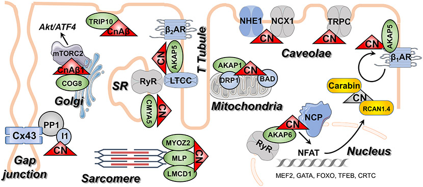
Calcineurin, also known as PP2B or PPP3, is a member of the PPP family of protein phosphatases that also includes PP1 and PP2A. Together these three phosphatases carryout the majority of dephosphorylation events in the heart. Calcineurin is distinct in that it is activated by the binding of calcium/calmodulin (Ca2+/CaM) and therefore acts as a node for integrating Ca2+ signals with changes in phosphorylation, two fundamental intracellular signaling cascades. In the heart, calcineurin is primarily thought of in the context of pathological cardiac remodeling, acting through the Nuclear Factor of Activated T-cell (NFAT) family of transcription factors. However, calcineurin activity is also essential for normal heart development and homeostasis in the adult heart. Furthermore, it is clear that NFAT-driven changes in transcription are not the only relevant processes initiated by calcineurin in the setting of pathological remodeling. There is a growing appreciation for the diversity of calcineurin substrates that can impact cardiac function as well as the diversity of mechanisms for targeting calcineurin to specific sub-cellular domains in cardiomyocytes and other cardiac cell types. Here, we will review the basics of calcineurin structure, regulation, and function in the context of cardiac biology. Particular attention will be given to: the development of improved tools to identify and validate new calcineurin substrates; recent studies identifying new calcineurin isoforms with unique properties and targeting mechanisms; and the role of calcineurin in cardiac development and regeneration.
Keywords: Calcineurin; Calcium signaling; Cardiac development; Heart failure; Hypertrophy; Intracellular signaling.
Copyright © 2021 Elsevier Inc. All rights reserved.
Figures


Similar articles
-
Cardiac anti-remodelling effect of aerobic training is associated with a reduction in the calcineurin/NFAT signalling pathway in heart failure mice.J Physiol. 2009 Aug 1;587(Pt 15):3899-910. doi: 10.1113/jphysiol.2009.173948. Epub 2009 Jun 8. J Physiol. 2009. PMID: 19505981 Free PMC article.
-
Calcineurin/NFAT coupling participates in pathological, but not physiological, cardiac hypertrophy.Circ Res. 2004 Jan 9;94(1):110-8. doi: 10.1161/01.RES.0000109415.17511.18. Epub 2003 Dec 1. Circ Res. 2004. PMID: 14656927
-
PICOT attenuates cardiac hypertrophy by disrupting calcineurin-NFAT signaling.Circ Res. 2008 Mar 28;102(6):711-9. doi: 10.1161/CIRCRESAHA.107.165985. Epub 2008 Feb 7. Circ Res. 2008. PMID: 18258855
-
Calcineurin-NFAT signaling regulates the cardiac hypertrophic response in coordination with the MAPKs.Cardiovasc Res. 2004 Aug 15;63(3):467-75. doi: 10.1016/j.cardiores.2004.01.021. Cardiovasc Res. 2004. PMID: 15276472 Review.
-
Calcium-calcineurin signaling in the regulation of cardiac hypertrophy.Biochem Biophys Res Commun. 2004 Oct 1;322(4):1178-91. doi: 10.1016/j.bbrc.2004.07.121. Biochem Biophys Res Commun. 2004. PMID: 15336966 Review.
Cited by
-
The Role of Interventional Irisin on Heart Molecular Physiology.Pharmaceuticals (Basel). 2022 Jul 14;15(7):863. doi: 10.3390/ph15070863. Pharmaceuticals (Basel). 2022. PMID: 35890161 Free PMC article.
-
Inhibition of adenylyl cyclase 8 prevents the upregulation of Orai1 channel, which improves cardiac function after myocardial infarction.Mol Ther. 2024 Mar 6;32(3):646-662. doi: 10.1016/j.ymthe.2024.01.026. Epub 2024 Jan 29. Mol Ther. 2024. PMID: 38291755 Free PMC article.
-
Betulinic Acid Improves Cardiac-Renal Dysfunction Caused by Hypertrophy through Calcineurin-NFATc3 Signaling.Nutrients. 2021 Sep 30;13(10):3484. doi: 10.3390/nu13103484. Nutrients. 2021. PMID: 34684485 Free PMC article.
-
Combined Pharmacological Modulation of Translational and Transcriptional Activity Signaling Pathways as a Promising Therapeutic Approach in Children with Myocardial Changes.Biomolecules. 2024 Apr 13;14(4):477. doi: 10.3390/biom14040477. Biomolecules. 2024. PMID: 38672493 Free PMC article. Review.
-
The Microenvironment of the Pathogenesis of Cardiac Hypertrophy.Cells. 2023 Jul 4;12(13):1780. doi: 10.3390/cells12131780. Cells. 2023. PMID: 37443814 Free PMC article. Review.
References
-
- Stewart AA, Ingebritsen TS, Manalan A, Klee CB, Cohen P, Discovery of a Ca2+- and calmodulin-dependent protein phosphatase: probable identity with calcineurin (CaM-BP80), FEBS Lett 137(1) (1982) 80–4. - PubMed
-
- Manalan AS, Klee CB, Affinity selection of chemically modified proteins: role of lysyl residues in the binding of calmodulin to calcineurin, Biochemistry 26(5) (1987) 1382–90. - PubMed
-
- Sussman MA, Lim HW, Gude N, Taigen T, Olson EN, Robbins J, Colbert MC, Gualberto A, Wieczorek DF, Molkentin JD, Prevention of cardiac hypertrophy in mice by calcineurin inhibition, Science 281(5383) (1998) 1690–3. - PubMed
Publication types
MeSH terms
Substances
Grants and funding
LinkOut - more resources
Full Text Sources
Miscellaneous
