. 2021 Nov:123:104705.
doi: 10.1016/j.jmbbm.2021.104705. Epub 2021 Aug 14.

Regional biomechanical and failure properties of healthy human ascending aorta and root

Affiliations

Regional biomechanical and failure properties of healthy human ascending aorta and root

Yue Xuan et al. J Mech Behav Biomed Mater. 2021 Nov.

Abstract

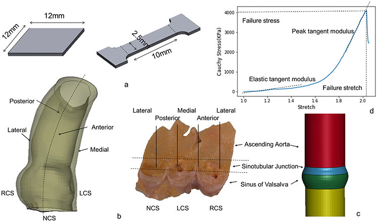
Purpose: Aortic dissection (AD) is a life-threatening event that occurs when the intimal entry tear propagates and separates inner from outer layers of the aorta. Diameter, the current criterion for aneurysm repair, is far from ideal and additional evidence to optimize clinical decision would be extremely beneficial. Biomechanical investigation of the regional failure properties of aortic tissue is essential to understand and proactively prevent AD. We previously studied biaxial mechanical properties of healthy human aorta. In this study, we investigated the regional failure properties of healthy human ascending aorta (AscAo) including sinuses of Valsalva (SOV), and sinotubular junction (STJ).

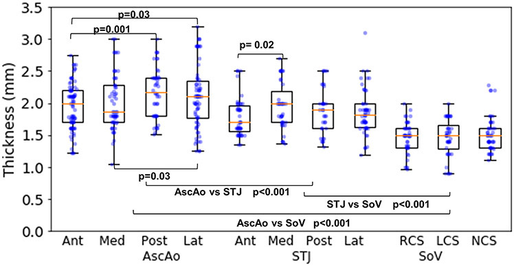
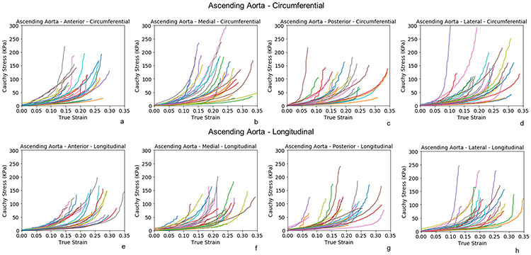
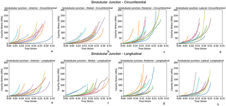
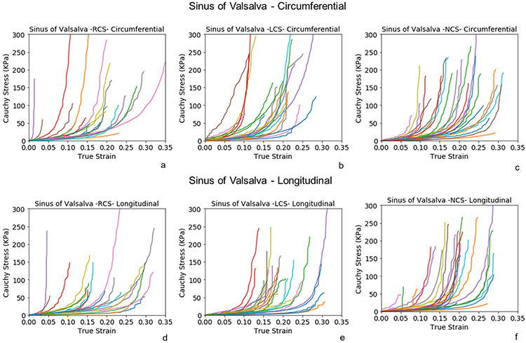
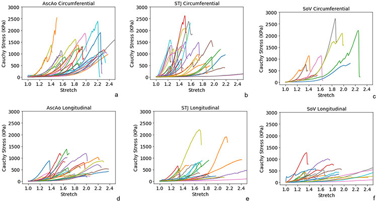
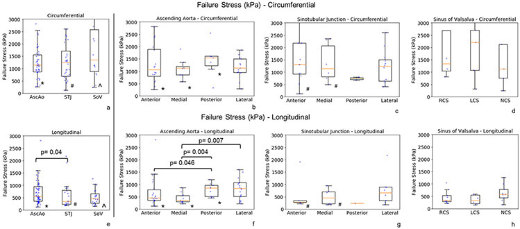
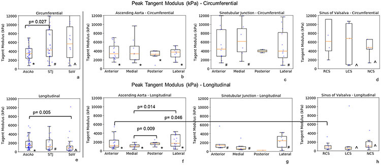
Results: A total of 430 intact tissue samples were harvested from 19 healthy donors whose hearts were excluded from heart transplantation. The donors had mean age of 51 ± 11.7 years and nearly equal gender distribution. Samples were excised from aortic regions and subregions at defined locations. Tissue strips were subjected to either biaxial or uniaxial failure testing. Wall thickness varied regionally being thickest at AscAo (2.08 ± 0.66 mm) and thinnest at SOV (1.46 ± 0.31 mm). Biaxial testing demonstrated hyperplastic behavior of aortic tissues. Posterior and lateral STJ subregions were found to be stiffer than anterior and medial subregions in both circumferential and longitudinal directions. Failure stresses were significantly higher in the circumferential than longitudinal directions in each subregion of AscAo, STJ, and SOV. Longitudinal failure stresses were significantly greater in AscAo than those in STJ or SOV. Longitudinal failure stresses in AscAo were much smaller anteriorly than posteriorly, and medially than laterally.

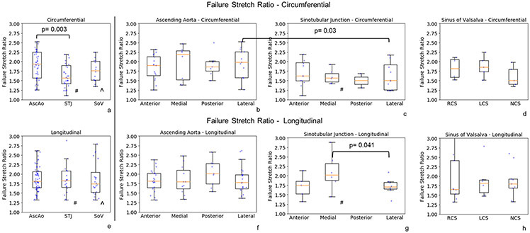
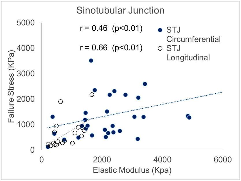
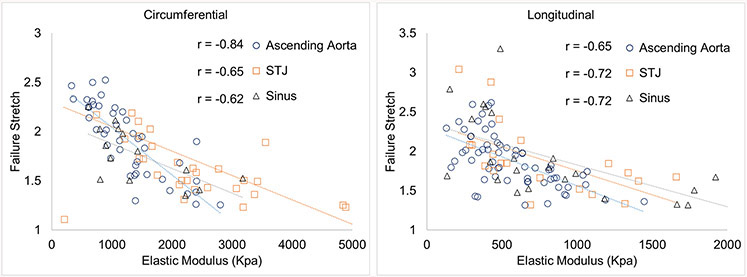
Conclusions: The finding of weakest region at the sinotubular junction along the longitudinal direction corroborates clinical observations of that region being commonly involved as the initial site of intimal tear in aortic dissections. Failure stretch ratios correlated to elastic modulus at each region. Furthermore, strong correlation was seen between STJ failure stresses and elastic modulus at physiological pressure along both circumferential and longitudinal directions. Correlating in-vivo aortic elastic modulus based on in-vivo imaging with experimentally determined elastic modulus at physiological pressure and failure stresses may potentially provide valuable information regarding aortic wall strength. Better understanding of aortic biomechanics in normal physiologic and aneurysmal pathologic states may aid in determining risk factors for predicting dissection in patient-specific fashion.

Keywords: Aortic dissection; Ascending aorta; Biaxial; Biomechanics; Failure stress; Healthy; Peak tangent modulus; Stretch.


Conflict of interest statement

Conflict of Interest Statement: The authors have no conflicts of interest to declare.

Figures

Figure 1a.
Figure 1a.
Sketch of biaxial and failure sample dimensions. 1b. Subregions of ascending aorta and aortic root. 1c. Sketch of the simplified model with distinct diameter and thickness. 1d. Representative plot of failure stress vs stretch. Dotted lines are labeled to describe the point where failure stress and stretch are measured. In addition, two dotted lines are labeled to describe the elastic and peak tangent moduli.
Figure 2:
Figure 2:
Thickness in regions and subregions of aortic root and ascending aorta.
Figure 3:
Figure 3:
Biaxial stress-strain curves of subregions of healthy ascending aorta along the circumferential(a-d) and longitudinal directions (e-h).
Figure 4:
Figure 4:
Biaxial stress-strain curves of the subregions of healthy sinotubular junction along the circumferential(a-d) and longitudinal directions (e-h).
Figure 5:
Figure 5:
Biaxial stress-strain curves of the subregions of healthy sinuses of Valsalva along the circumferential(a-c) and longitudinal directions (d-f).
Figure 6:
Figure 6:
Representative failure behavior at regions of aortic root and ascending aorta along the circumferential (a-c) and longitudinal (d-f) directions.
Figure 7:
Figure 7:
Failure stresses at regions (a, e) and subregions (b-d, f-h) of aortic root and ascending aorta along the circumferential(a-d) and longitudinal directions (e-h). The raw data (dots) were overlaid on the median and (25%, 75%) interquartile range. * denotes the statistically significant differences in ascending aorta between circumferential (circ) and longitudinal (long) directions - AscAo circ vs long, p=0.001; AscAo anterior circ vs long, p=0.003; AscAo medial circ vs long, p<0.001; AscAo posterior circ vs long, p=0.013. # denotes the statistically significant differences in sinotubular junction between circumferential and longitudinal directions - STJ circ vs long, p<0.001; STJ anterior circ vs long, p=0.031; STJ medial circ vs long, p=0.015. ^ denotes the statistically significant differences in sinus of Valsalva between circumferential and longitudinal directions - SOV circ vs long, p<0.001.
Figure 8:
Figure 8:
Failure stretches at regions (a, e) and subregions (b-d, f-h) of aortic root and ascending aorta along the circumferential(a-d) and longitudinal directions (e-h). The raw data (dots) were overlaid on the median and (25%, 75%) interquartile range. The raw data (dots) were overlaid on the median and (25%, 75%) interquartile range. # denotes the statistically significant differences in sinotubular junction between circumferential and longitudinal directions - STJ circ vs long, p=0.034; STJ medial circ vs long, p=0.023. ^ denotes the statistically significant differences in sinus of Valsalva between circumferential and longitudinal directions - SOV circ vs long, p=0.048.
Figure 9:
Figure 9:
Peak tangent moduli at regions (a, e) and subregions (b-d, f-h) of aortic root and ascending aorta along the circumferential(a-d) and longitudinal directions (e-h). The raw data (dots) were overlaid on the median and (25%, 75%) interquartile range. In * denotes the statistically significant differences in ascending aorta between circumferential (circ) and longitudinal (long) directions - AscAo circ vs long, p=0.001; AscAo anterior circ vs long, p<0.001; AscAo medial circ vs long, p<0.001; AscAo posterior circ vs long p=0.01; AscAo lateral circ vs long p=0.037. # denotes the statistically significant differences in sinotubular junction between circumferential and longitudinal directions - STJ circ vs long, p<0.001; STJ anterior circ vs long, p=0.015; STJ medial circ vs long p=0.004; STJ lateral circ vs long, p=0.041. λ denotes the statistically significant differences in sinus of Valsalva between circumferential and longitudinal directions - SOV cir vs long, p<0.001; SOV left coronary sinus circ vs long p=0.042; SOV non-coronary sinus circ vs long, p=0.008.
Figure 10.
Figure 10.
Correlation between failure stresses and relative elastic tangent modulus at sinotubular junction along the circumferential and longitudinal directions.
Figure 11.
Figure 11.
Correlation between failure stretch ratio and elastic tangent modulus at regions of aortic root and ascending aorta along the circumferential and longitudinal directions.

References

    1. Azadani AN, Chitsaz S, Mannion A, Mookhoek A, Wisneski A, Guccione JM, Hope MD, Ge L, Tseng EE, 2013. Biomechanical properties of human ascending thoracic aortic aneurysms. Ann Thorac Surg 96, 50–58. - PubMed
    1. Azadani AN, Chitsaz S, Matthews PB, Jaussaud N, Leung J, Tsinman T, Ge L, Tseng EE, 2012a. Comparison of mechanical properties of human ascending aorta and aortic sinuses. Ann Thorac Surg 93, 87–94. - PubMed
    1. Azadani AN, Chitsaz S, Matthews PB, Jaussaud N, Leung J, Wisneski A, Ge L, Tseng EE, 2012b. Biomechanical comparison of human pulmonary and aortic roots. European journal of cardio-thoracic surgery : official journal of the European Association for Cardio-thoracic Surgery 41, 1111–1116. - PubMed
    1. Cacho F, Elbischger PJ, Rodríguez JF, Doblaré M, Holzapfel GA, 2007. A constitutive model for fibrous tissues considering collagen fiber crimp. International Journal of Non-Linear Mechanics 42, 391–402.
    1. Choudhury N, Bouchot O, Rouleau L, Tremblay D, Cartier R, Butany J, Mongrain R, Leask RL, 2009. Local mechanical and structural properties of healthy and diseased human ascending aorta tissue. Cardiovascular Pathology 18, 83–91. - PubMed

Publication types