. 2021 Oct:56:102507.
doi: 10.1016/j.scr.2021.102507. Epub 2021 Aug 19.

Establishment of human induced trophoblast stem-like cells from term villous cytotrophoblasts

Affiliations

Establishment of human induced trophoblast stem-like cells from term villous cytotrophoblasts

Tao Bai et al. Stem Cell Res. 2021 Oct.

Abstract

Human trophoblast stem cells (hTSC) can be isolated from first trimester placenta but not from term placenta. Here we demonstrate that villous cytotrophoblasts (vCTB) from term placenta can be reprogrammed into induced trophoblastic stem-like cells (iTSC) by introducing sets of transcription factors. The iTSCs express TSC markers such as GATA3, TEAD4 and ELF5, and are multipotent, validated by their differentiation into both extravillous trophoblasts (EVT) and syncytiotrophoblasts (STB) in vitro and in vivo. The iTSC can be passaged indefinitely in vitro without slowing of growth. The transcriptome profile of these cells closely resembles the profile of hTSC isolated from first trimester placentae but different from the term placental vCTB from which they originated. The ability to reprogram cells from term placenta into iTSC will allow study of early gestation events which impact placental function later in gestation, including preeclampsia and spontaneous preterm birth.

Keywords: Differentiation; Placenta; Reprogramming; Stem cell; Trophoblast.


Conflict of interest statement

Declaration of Competing Interest

The authors declare that they have no known competing financial interests or personal relationships that could have appeared to influence the work reported in this paper.

Figures

Fig. 1.
Fig. 1.
Reprogramming term placental villous cytotrophoblasts to iTSC. (A) Schematic representation of protocols to reprogram term vCTB to iTSC. (B) Mixtures of 5–6 transcription factors (TFs) were used to induce reprogramming of term vCTB. Isolated vCTB from term placental cores were transduced under the indicated combinations of TFs for 1 day. The transduced cells were switched to reprogramming medium 24 h after removal of virus mixtures. The signs of + indicate included transcription factors. (C) Immunofluorescent images of vCTB prior to viral transduction stained with epithelial marker cytokeratin 7 (CK7), stromal marker Vimentin (Vim), and cell cycle marker Ki67. (C’-E) Phase-contrast images of cells from well 2 (5 factors minus GATA3) at day 1 (D1), day 14 (D14), and after 10 passages (P10) showing an epithelial-like colony emerging within the differentiated STB sheet around day 14–21 (D), and remain morphologically similar and proliferative at passage 10 (E). Scale bar, C = 100 μm; D-F = 500 μm.
Fig. 2.
Fig. 2.
Detection of hTSC markers in iTSC and STB and EVT markers after iTSC differentiation. (A-J) Immunostaining for TSC markers GATA3, TFAP2C, TEAD4, ELF5, ITGA6, TP63, CDH1, CK7 and Ki67 (Green) in 5F-G3 iTSC at passage 10–15. Nuclei were stained with DAPI (Blue). Similar results were obtained with three independent cell lines generated using the six factors (sup Fig. 1). (K, K’) Immunostaining of STB marker hCG (green) in iTSC cultured in TSC medium (K) or STB differentiation medium (K’) at 3 days in vitro demonstrating the capacity of iTSC to generate of multinucleated syncytiotrophoblasts under appropriate differentiation conditions. Similar results were obtained with two independent cell lines. (L, L’) Immunostaining of HLA-G (green) in iTSC cultured in TSC medium (L) or EVT differentiation medium (L’) at 7 days in vitro demonstrating the capacity of iTSC to generate HLA-G+ cells with bipolar morphology under appropriate differentiation conditions. Similar results were obtained with two independent cell lines. (M-N’) Representative images of CK7 (M’) and HLA-G (N’) expressing cells detected on the bottom surface of the Transwell membrane after iTSC cultured in TSC medium (M, N) or EVT medium (M’, N’) on the top surface of Transwell membrane for 7 days. Only iTSC cells in EVT differentiation medium penetrated Transwells membrane. scale bar = 100 μm.
Fig. 3.
Fig. 3.
RNA-Sequencing revealed transcriptome level similarities between iTSC and hTSC. (A) Principle component analysis of hTSC, iTSC and untreated term cytotrophoblasts, iTSC and hTSC aligned closely on the PC1 and PC2 axes but showed variability on the PC3 axis. All data points were collected with three technical triplicates. (B) Expression comparisons of proliferative cytotrophoblast-related genes between vCTB, hTSC and iTSC revealed iTSCs show similar expression profiles with hTSC and significant differences from vCTB. (C) Expression levels of proliferative cytotrophoblast-related genes in primary and reprogrammed iTSC cells. Data are presented as mean + SD. n = 3 technical triplicates. (D) Quantitative real-time PCR analysis of proliferative cytotrophoblast-related gene expression in vCTB (n = 3) and iTSC (n = 3). The transcript level of TEAD4 was higher (2.16 fold increase, p < 0.01) in iTSC than in vCTB. Similarly, ELF5 expression in elevated in iTSC compared to vCTB (7.6 fold increase, p < 0.001. The data were normalized to GAPDH expression and represented as fold change relative to vCTB. ** p < 0.01; ***p < 0.001.
Fig. 4.
Fig. 4.
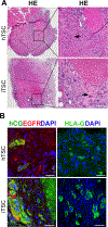
Modified teratoma assays demonstrate differentiation of iTSC into STB and EVT lineages in vivo. (A) Hematoxylin-eosin (HE) staining in hTSC and iTSC-derived lesion images at 4X magnification (right panel) with 10X magnification view of the lesion boarder (right panel). Some STB-like trophoblast cells contained blood-filled lacunae (arrows). (B) Immunofluorescence images of hTSC and iTSC-derived lesions. Differentiation into STB and EVT in the lesion was demonstrated by immunostaining of EGFR1 (red) and hCG (green) for STB and for HLA-G (green) for EVT with nuclei stained with DAPI. Scale bar, A = 100 μm, B = 50 μm.

References

    1. Chu Cheung V, Peng CY, Marinic M, Sakabe NJ, Aneas I, Lynch VJ, Ober C, Nobrega MA, Kessler JA, 2021. Pluripotent cell cell-derived endometrial stromal fibroblasts in a cyclic, hormone-responsive, coculture model of human decidua. Cell Rep. 35(7):109138. - PubMed
    1. Cinkornpumin JK, Kwon SY, Guo Y, Hossain I, Sirois J, Russett CS, Tseng H-W, Okae H, Arima T, Duchaine TF, Liu W, Pastor WA, 2020. Naive human embryonic stem cells can give rise to cells with a trophoblast-like transcriptome and methylome. Stem Cell Rep. 15 (1), 198–213. - PMC - PubMed
    1. Coorens THH, Oliver TRW, Sanghvi R, Sovio U, Cook E, Vento-Tormo R, Haniffa M, Young MD, Rahbari R, Sebire N, Campbell PJ, Charnock-Jones DS, Smith GCS, Behjati S, 2021. Inherent mosaicism and extensive mutation of human placentas. Nature 592 (7852), 80–85. - PMC - PubMed
    1. Dong C, Beltcheva M, Gontarz P, Zhang B, Popli P, Fischer LA, Khan SA, Park KM, Yoon EJ, Xing X, Kommagani R, Wang T, Solnica-Krezel L, Theunissen TW, 2020. Derivation of trophoblast stem cells from naïve human pluripotent stem cells. Elife. 12 (9), e52504. - PMC - PubMed
    1. Guo G.e., Stirparo GG, Strawbridge SE, Spindlow D, Yang J, Clarke J, Dattani A, Yanagida A, Li MA, Myers S, Özel BN, Nichols J, Smith A, 2021. Human naive epiblast cells possess unrestricted lineage potential. Cell Stem Cell 28 (6), 1040–1056.e6. - PMC - PubMed

Publication types