. 2021 Dec:346:113845.
doi: 10.1016/j.expneurol.2021.113845. Epub 2021 Aug 26.

Astrocyte inflammatory signaling mediates α-synuclein aggregation and dopaminergic neuronal loss following viral encephalitis

Affiliations

Astrocyte inflammatory signaling mediates α-synuclein aggregation and dopaminergic neuronal loss following viral encephalitis

Collin M Bantle et al. Exp Neurol. 2021 Dec.

Abstract

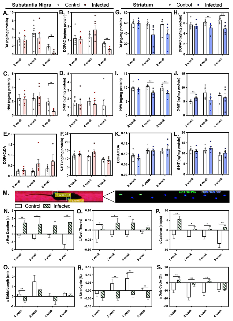
Viral infection of the central nervous system (CNS) can cause lasting neurological decline in surviving patients and can present with symptoms resembling Parkinson's disease (PD). The mechanisms underlying postencephalitic parkinsonism remain unclear but are thought to involve increased innate inflammatory signaling in glial cells, resulting in persistent neuroinflammation. We therefore studied the role of glial cells in regulating neuropathology in postencephalitic parkinsonism by studying the involvement of astrocytes in loss of dopaminergic neurons and aggregation of α-synuclein protein following infection with western equine encephalitis virus (WEEV). Infections were conducted in both wildtype mice and in transgenic mice lacking NFκB inflammatory signaling in astrocytes. For 2 months following WEEV infection, we analyzed glial activation, neuronal loss and protein aggregation across multiple brain regions, including the substantia nigra pars compacta (SNpc). These data revealed that WEEV induces loss of SNpc dopaminergic neurons, persistent activation of microglia and astrocytes that precipitates widespread aggregation of α-synuclein in the brain of C57BL/6 mice. Microgliosis and macrophage infiltration occurred prior to activation of astrocytes and was followed by opsonization of ⍺-synuclein protein aggregates in the cortex, hippocampus and midbrain by the complement protein, C3. Astrocyte-specific NFκB knockout mice had reduced gliosis, α-synuclein aggregate formation and neuronal loss. These data suggest that astrocytes play a critical role in initiating PD-like pathology following encephalitic infection with WEEV through innate immune inflammatory pathways that damage dopaminergic neurons, possibly by hindering clearance of ⍺-synuclein aggregates. Inhibiting glial inflammatory responses could therefore represent a potential therapy strategy for viral parkinsonism.

Keywords: Alpha-synuclein; Alphaviruses; Glia; Neurodegeneration; Neuroinflammation; Parkinson’s Disease; Viral encephalitis; Western equine encephalitis virus.


Conflict of interest statement

Competing Interest

The authors declare no competing interest

Competing interests

The authors declare that there exist no competing interests or conflicts, disclosed or otherwise. All research and data will be made freely accessible, per the guidelines of the National Institutes of Health.

Collin M. Bantle

Savannah M. Rocha

C. Tenley French

Aaron T. Phillips

Kevin Tran

Kenneth E. Olson

Todd A. Bass

Tawfik Aboellail

Richard J. Smeyne

Ronald B. Tjalkens

Figures

Figure 1.
Figure 1.. Intranasal inoculation with recombinant WEEV coupled with immunotherapy facilitates viral propagation and persistent infection throughout the CNS without incapacitating the mice.
(A) Schematic illustration of recombinant viral sequence expressing dsRed used and the associated treatment scheme. SPG - subgenomic promoter internal initiation site, UTR - untranslated region, DsRed – destabilized red fluorescent protein. Intranasal inoculation results in a persistent infection, with WEEV replicating in neurons and not in glia in the CNS. (B - D) Entire sagittal sections of mouse brain were imaged by digital montaging following intranasal inoculation with DsRed-expressing WEEV (1×10⁴ PFU/ml) and co-immunostained for the astrocyte marker, glial fibrillary acidic protein GFAP (B), the microglia marker, ionized calcium binding adaptor molecule IBA1 (C) or the dopaminergic neuronal marker, tyrosine hydroxylase TH (D). High resolution insets depict cellular staining in the substantia nigra, hippocampus and olfactory bulb. Coronal sections (E - G) were imaged separately, with high resolution insets depicting region-specific cellular staining with dsRed and GFAP, IBA1 and TH (1-3). (H) Schematic illustration of recombinant viral sequence expressing firefly luciferase used and the associated treatment scheme. Pseudo-colored images of luciferase-activity following infection with WEEV-Luc were collected at Ohr (I), 12hr PI (J), 48hr PI (K) and 8WPI (L). (M) Following intranasal infection with WEEV-Luc, mice were treated with anti-E1 passive immunotherapy at 12hr and 48hr post-infection to maintain a consistent infection within established level of total lumen flux (red lines). (N) Survival curves for control mice (blue symbols), mice receiving I.N. WEEV + immunotherapy (green symbols) and mice receiving I.N. WEEV without immunotherapy (red symbols).
Figure 2.
Figure 2.. Intranasal infection with WEEV causes dopaminergic neuronal loss, microgliosis and invastion of peripheral macrophages in the substantia nigra pars compacta.
(A-H) Representative IHC images of the substantia nigra pars compacta (SNpc) from mice infected or mock-infected with saline at 1,2,4, and 8WPI. (I) Stereological assessment of dopaminergic neurons in the SNpc. (J) Stereological assessment of microglia in the SN. (K-M) Quantification of resting and active microglia and infiltrating peripheral macrophages. (N-P) Linear dot-plot representation of cell counts following encephalitic infection with WEEV at 1,2,4, and 8WPI. (Q) Normalized cell count overlay of dopaminergic neurons (green), microgliosis (grey), and macrophage infiltration (blue) at 1, 2, and 8WPI. (*p<0.05, ** p<0.005, ***p<0.0005, n=6-8 per group)
Figure 3.
Figure 3.. Encephalitic infection with WEEV alters catecholamine homeostasis in the brain with associated neurobehavioral abnormalities in C57Bl/6 mice.
Levels of catecholamines and associated metabolites were measured in substantia nigra and striatum by high performance liquid chromatography (HPLC) analysis and electrochemical detection. (A, G) Dopamine (DA) levels in the substantia nigra (SN) and striatum (ST), respectively, along with DA metabolites 3,4-dihydroxyphenyl-acetic acid (DOPAC) (B, H), homovanillic acid (HVA) (C, I), and 3-methoxytyramine (3MT) (D, J) were measured at 2, 4 and 8WPI, as well as the DOPAC:DA ratio (E, K). Levels of serotonin (5-HT) (F, L) were also determined at 2, 4, and 8WPI in each brain region. (M) The gait and locomotor function of freely moving animals was analyzed at 1, 2, 4 and 8WPI. Parameters evaluated included run duration (N), rest time (O), cadence (P), stride length (Q), step cycle (R) and duty cycle (S) and were used to identify neurological changes associated with WEEV infection. (*p<0.05, ** p<0.005, ***p<0.0005, n=4-8 per group)
Figure 4.
Figure 4.. Encephalitic infection with WEEV induces neuroinflammatory activation of astrocytes in the SNpc.
(A-H) Representative 10X montage immunofluorescence images of control and infectd mice at 1, 2, 4 and 8 WPI. Sections were co-immunostained with S100β (cyan), anti-C3 (red), and DAPI (blue) with 40X high magnification insets of the SNpc. (I) Quantification of S100β + astrocytes in the SNpc at 1,2,4, and 8 WPI. (J) Quantitative analysis of C3 intensity within astrocyte-specific regions of interest (ROIs) localized to the SNpc in control and infected animals at all time points. (K) Time-dependent determination of C3 levels in S100β + astrocytes spanning all time-points in infected animals. (*p<0.05, ** p<0.005, ***p<0.0005, n=6-8 per group)
Figure 5.
Figure 5.. WEEV induces rapid formation of α-synuclein protein plaques in the cortex, hippocampus and midbrain of surviving wild-type mice.
Representative images and pathological scoring of P129+ immunohistochemical staining from mice infected for 1WPI (A-E), 2WPI (F-J), 4WPI (K-O) and 8WPI (P-T) with representative high magnification 40X inset images. The average pathological score in the cortex, hippocampus and midbrain at each timepoint is shown is black (A,F,K,P) as well as in each image panel. (*p<0.05, ** p<0.005, ***p<0.0005, n=6-8 per group). Brain sections were immunolabeled for expression of phospho-Eif2 (U,V) and phospho-PERK (W,X) and imaged by fluorescence micrscopy.
Figure 6.
Figure 6.. Encephalitic infection with WEEV induces opsonization of α-synuclein protein aggregates in the cortex, hippocampus and midbrain with astrocyte-derived complement C3 protein.
Immunofluorescence images of complement C3b (red) co-localization with P129+ protein aggregates (green) were analyzed in the cortex (A-C), hippocampus (D-F) and midbrain (G-I) at various times following infection with WEEV. Nuclei were counterstained with DAPI (blue). High magnification 40X inset images are depicted from each region. Representative images panels are presented from 2 WPI, the time of maximal P129 aggregation. (J) Quantification of C3b deposition co-localizing with protein aggregates within each brain region was determined at 2, 4 and 8WPI by analyzing the amount of C3b co-localizing with regions of interest detecting P129+ aggregates. (*p<0.05, **p<0.005, ***p<0.0005, n=6-8 per group)
Figure 7.
Figure 7.. Genetic knockout of NFκB in astrocytes reduces gliosis and α-synuclein aggregation throughout the brain.
(A) Schematic illustration of the Cre-recombinase under control of the human glial fibrillary acidic protein promoter (hGFAP). Hemizygous GFAP-Cre mice were crossed with I kappa B kinase 2 (Ikk2)-loxP mice, which facilitated selective deletion of IKK2 in astrocytes and provided a cell-specific knockout of NF-κB in astrocytes. hGFAP-cre+/−/IKK2fl/fl (KO) or hGFAP-cre−/−/IKK2fl/fl (WT) animals were intranasally infected with WEEV or mock-infected (control) with saline and treated with anti-E1immunotherapy. (B-D) IHC images with high magnification 40X inset images of P129+ staining from infected hGFAP-cre−/−/IKK2fl/fl WT mice, (E-G) uninfected control hGFAP-cre+/−/IKK2fl/fl KO mice and (H-J) infected hGFAP-cre+/−/IKK2fl/fl KO mice. (K) Pathological scoring of multiple brain regions was performed at 8WPI to quantify the extent of P129+ aggregation in each treatment group. (*p<0.05, **p<0.005, n=6-8 per group).
Figure 8.
Figure 8.. Microgliosis and peripheral macrophage infiltration precede astrogliosis and formation of phospho-Ser(129)-α-synuclein protein aggregates following intranasal infection with WEEV.
The kinetics of cellular responses to WEEV infection in the substantia nigra were modeled by curve fitting normalized data sets to generate representative plots of (A) monocyte infiltration and microglial activation, (B) astrocyte activation, (C) accumulation of P129+ protein aggregates and (D) dopaminergic neurodegeneration. (E) Combined overlays of each response during the 8-week course of infection. (F) Schematic representation of time-dependent cellular responses to WEEV infection indicated that glial activation and α-synuclein aggregation precede degeneration of dopaminergic neurons. (n=6-8 per group)

Similar articles

Cited by

References

    1. Alam G, et al. 2017. Single low doses of MPTP decrease tyrosine hydroxylase expression in the absence of overt neuron loss. Neurotoxicology. 60, 99–106. - PMC - PubMed
    1. Anderson JP, et al. 2006. Phosphorylation of Ser-129 is the dominant pathological modification of alpha-synuclein in familial and sporadic Lewy body disease. J Biol Chem. 281, 29739–52. - PubMed
    1. Bantle CM, et al. 2019. Infection with mosquito-borne alphavirus induces selective loss of dopaminergic neurons, neuroinflammation and widespread protein aggregation. NPJ Parkinsons Dis. 5, 20. - PMC - PubMed
    1. Bantle CM, et al. 2021. Manganese exposure in juvenile C57BL/6 mice increases glial inflammatory responses in the substantia nigra following infection with H1N1 influenza virus. PLoS One. 16, e0245171. - PMC - PubMed
    1. Baquet ZC, et al. 2009. A comparison of model-based (2D) and design-based (3D) stereological methods for estimating cell number in the substantia nigra pars compacta (SNpc) of the C57BL/6J mouse. Neuroscience. 161, 1082–90. - PMC - PubMed

Publication types

MeSH terms