. 2021 Nov:243:118520.
doi: 10.1016/j.neuroimage.2021.118520. Epub 2021 Aug 26.

Differential resting-state patterns across networks are spatially associated with Comt and Trmt2a gene expression patterns in a mouse model of 22q11.2 deletion

Affiliations

Differential resting-state patterns across networks are spatially associated with Comt and Trmt2a gene expression patterns in a mouse model of 22q11.2 deletion

Natalia Gass et al. Neuroimage. 2021 Nov.

Abstract

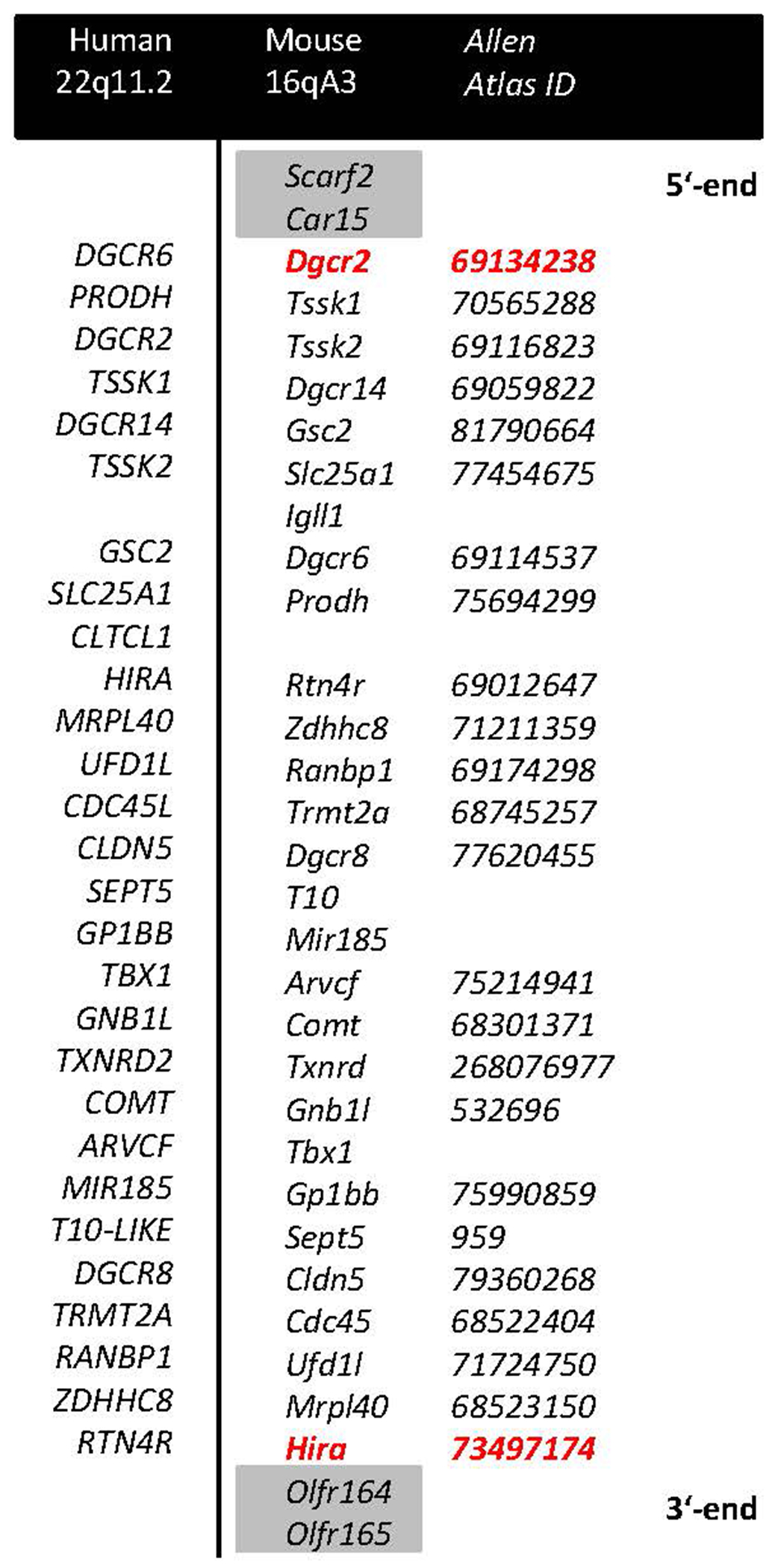
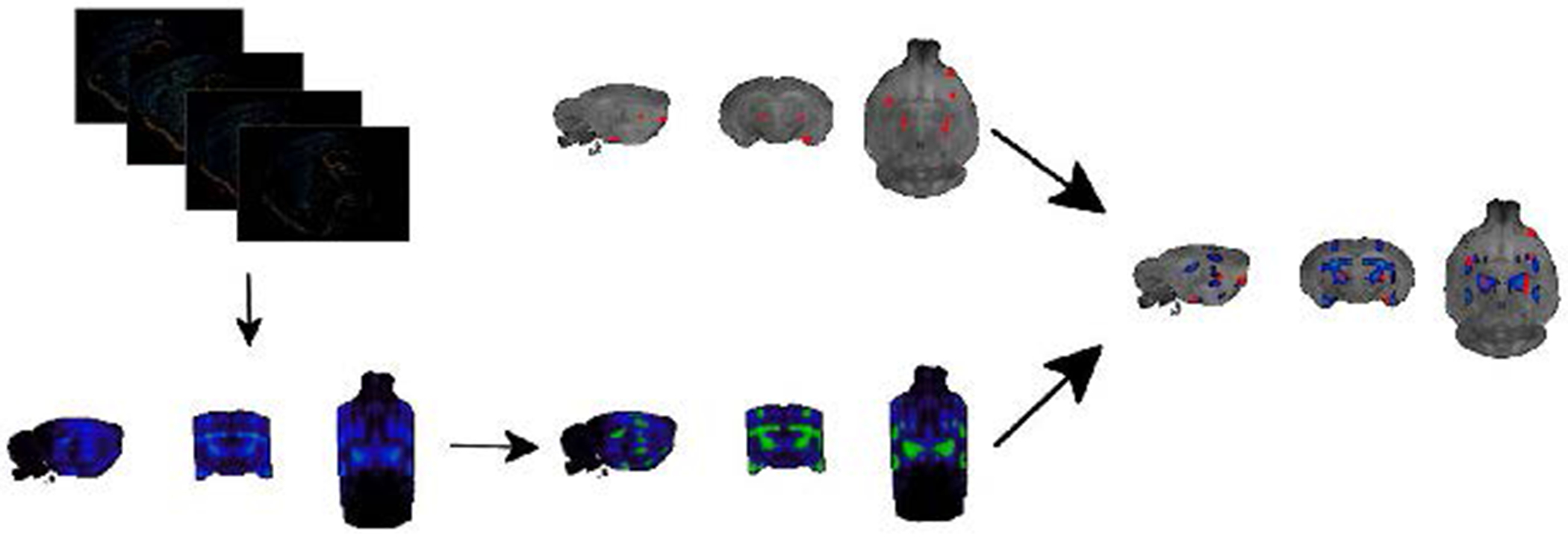
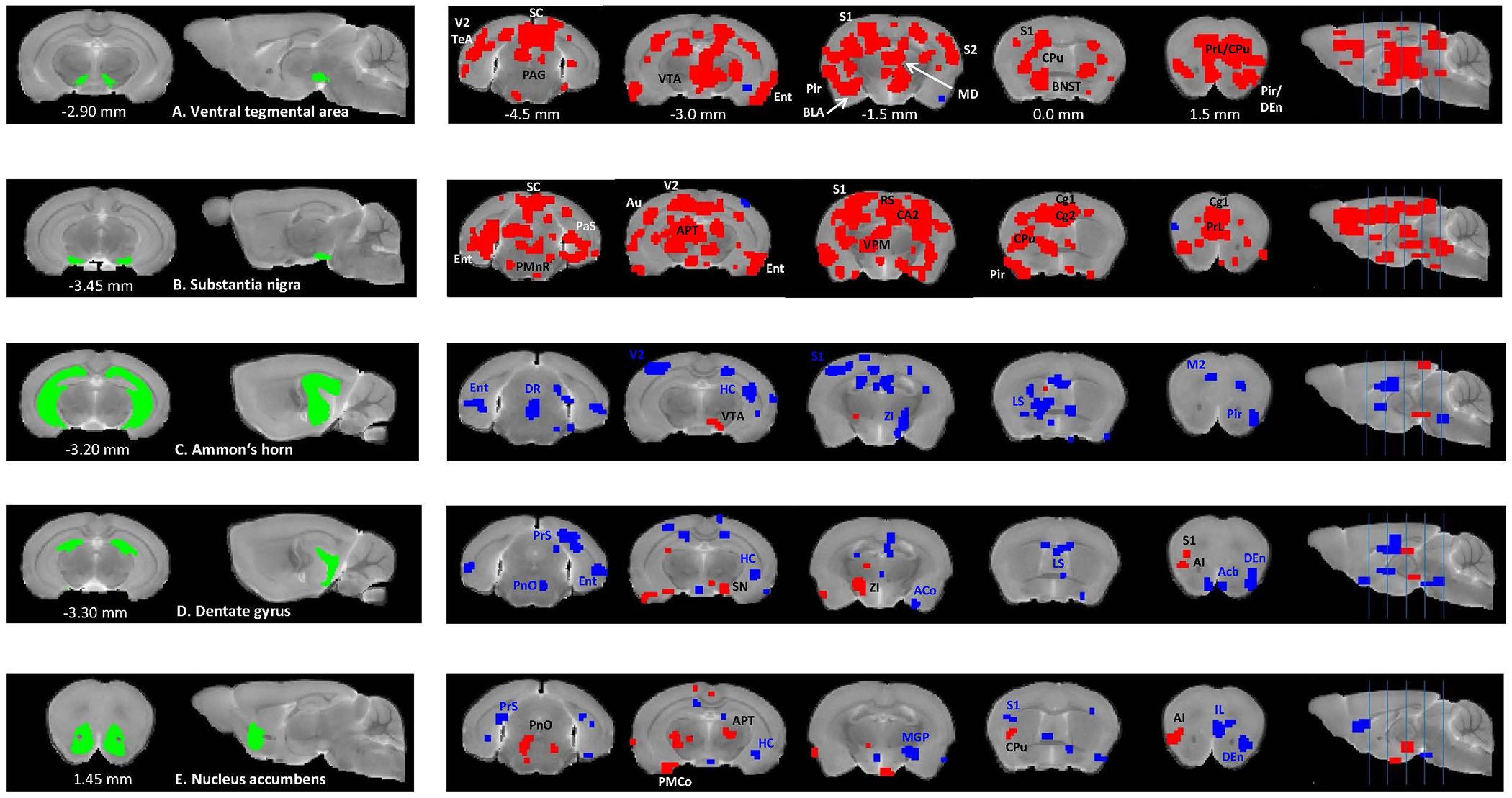
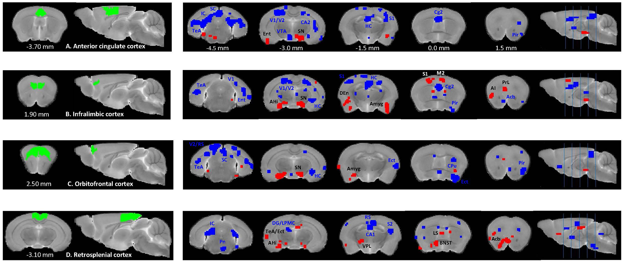
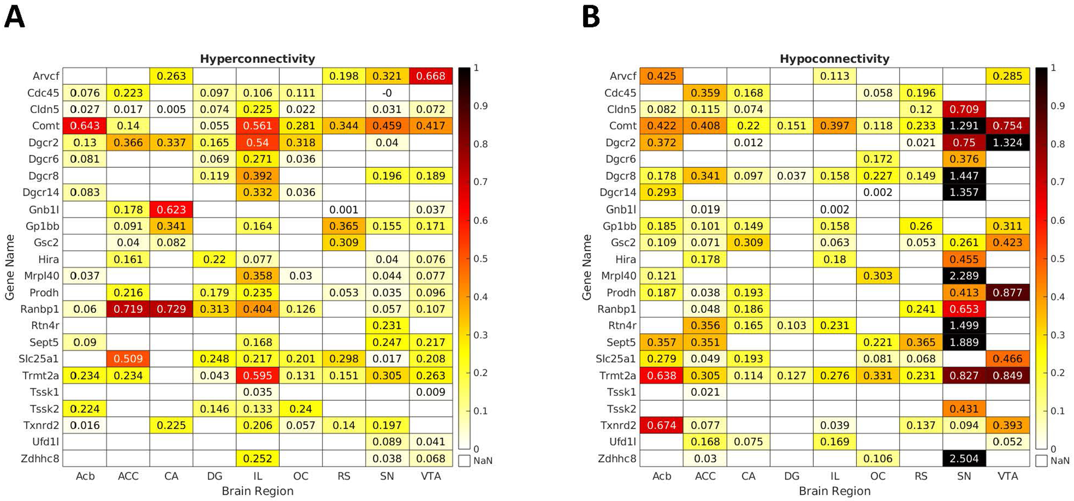
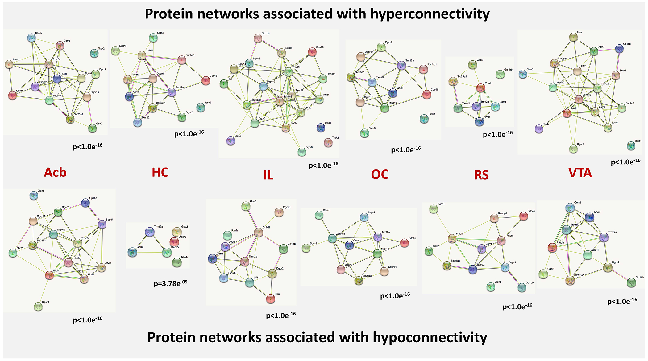
Copy number variations (CNV) involving multiple genes are ideal models to study polygenic neuropsychiatric disorders. Since 22q11.2 deletion is regarded as the most important single genetic risk factor for developing schizophrenia, characterizing the effects of this CNV on neural networks offers a unique avenue towards delineating polygenic interactions conferring risk for the disorder. We used a Df(h22q11)/+ mouse model of human 22q11.2 deletion to dissect gene expression patterns that would spatially overlap with differential resting-state functional connectivity (FC) patterns in this model (N = 12 Df(h22q11)/+ mice, N = 10 littermate controls). To confirm the translational relevance of our findings, we analyzed tissue samples from schizophrenia patients and healthy controls using machine learning to explore whether identified genes were co-expressed in humans. Additionally, we employed the STRING protein-protein interaction database to identify potential interactions between genes spatially associated with hypo- or hyper-FC. We found significant associations between differential resting-state connectivity and spatial gene expression patterns for both hypo- and hyper-FC. Two genes, Comt and Trmt2a, were consistently over-expressed across all networks. An analysis of human datasets pointed to a disrupted co-expression of these two genes in the brain in schizophrenia patients, but not in healthy controls. Our findings suggest that COMT and TRMT2A form a core genetic component implicated in differential resting-state connectivity patterns in the 22q11.2 deletion. A disruption of their co-expression in schizophrenia patients points out a prospective cause for the aberrance of brain networks communication in 22q11.2 deletion syndrome on a molecular level.

Keywords: 22q11.2 deletion; Comt; Functional connectivity; Mouse; Schizophrenia; Trmt2a; ventral tegmental area.


Figures

Figure 1.
Figure 1.. A schematic representation of deleted genes aligned for human (22q11.2) and mouse (16qA3) chromosomal regions.
Red color denotes genes targeted with a LoxP site, grey shade denotes genes at the border not comprising part of the deletion. IDs of the expression maps for the respective genes in the Allen Mouse Brain Atlas are reported in italics.
Figure 2.
Figure 2.. Overview over the pipeline for the joint analysis of gene expression patterns and differential resting state connectivity.
The top left image contains layered 2D expression images of the COMT gene which gets converted into the 3D image below. Please note that the lateral edges of the brain are not fully covered by the sagittal slices. Images then get binarized and aligned to the Waxholm space, which serves as a standard reference space for both gene expression maps and clusters indicating differential resting-state connectivity (top right image). After this alignment, a direct comparison between differential resting-state connectivity and gene expression patterns can be achieved.
Figure 3.
Figure 3.. Between-group comparisons of seed-based resting-state functional connectivity for the subcortical brain regions between Df(h22q11)/+ and wild type mice.
Two-sample t-tests (p <0.01) show significantly higher (red) and lower (blue) connectivity between ventral tegmental area (A), substantia nigra (B), Ammon’s horn (C), dentate gyrus (D), nucleus accumbens (E), and multiple cortical and subcortical brain regions in Df(h22q11)/+ mice, compared to the control group. Coordinates are in mm to Bregma. Abbreviations: ACo – anterior cortical amygdaloid nucleus, AI – agranular insular cortex, APT – anterior pretectal nucleus, Au – auditory cortex, BLA – basolateral amygdaloid nucleus, BNST – bed nucleus of stria terminalis, CA2 – field CA2 of the hippocampus, Cg1 – cingulate cortex area 1, Cg2 - cingulate cortex area 2, CPu – caudate putamen, DEn – dorsal endopiriform nucleus, DR – dorsal raphe nucleus, Ent – entorhinal cortex, HC - hippocampus, LS – lateral septal nucleus, M2 – secondary motor cortex, MD – mediodorsal thalamic nucleus, MGP – medial globus pallidus, PAG – periaqueductal gray, PaS - parasubiculum, Pir – piriform cortex, PMnR – paramedian raphe nucleus, PMCo – posteromedial cortical amygdaloid area, PnO – pontine reticular nucleus oral part, PRh – perirhinal cortex, PrL – prelimbic cortex, PrS - presubiculum, S1 – primary somatosensory cortex, S2 - secondary somatosensory cortex, SC – superior colliculus, TeA – temporal association cortex, V2 – secondary visual cortex, VPM – ventral posteromedial thalamic nucleus, ZI – zona incerta.
Figure 4.
Figure 4.. Between-group comparisons of seed-based rs-FC for the cortical brain regions between Df(h22q11)/+ and wild type mice.
Two-sample t-tests (p <0.01) show significantly higher (red) and lower (blue) connectivity between anterior cingulate cortex (A), infralimbic cortex (B), orbitofrontal cortex (C) and retrosplenial cortex (D) and multiple cortical and subcortical brain regions in Df(h22q11)/+ mice, compared to the control group. Coordinates are in mm to Bregma. Abbreviations: AHi – amygdalohippocampal area, Amyg - amygdala, Ect – ectorhinal cortex, LPMC – lateral posterior thalamic nucleus mediocaudal part, Pn – pontine nuclei, V1 – primary visual cortex, VPL – ventral posterolateral thalamic nucleus. Other abbreviations are the same as in Figure 3.
Figure 5.
Figure 5.. Genes overexpressed in networks indicating hyper- and hypoconnectivity for seed regions.
Depicted are the matrices presenting fold changes between neural networks hyper- (left) and hypoconnected (right) to the seed regions and spatial expression patterns of the hemi-deleted genes in these networks. Only values for significant overexpression are provided. Comt and Trmt2a were the most consistently implicated genes across the studied networks. Other genes showed expression patterns that were associated either with hyper- or hypoconnected networks.
Figure 6.
Figure 6.. Protein-protein interaction networks overexpressed in networks indicating hyper- and hypoconnectivity.
The nodes of the network represent the proteins, which are encoded by the genes entered into our analysis. The content of the node indicates, whether the 3D structure of the gene product is known or not. The colors of the edges indicate the kind of evidence for protein-protein interactions. Nodes (proteins) without edges indicate that a protein has no functional associations with other proteins of that contrast. The p-values under the networks indicate the likelihood that the involved genes form a protein-protein network above chance level. Known interactions: light blue – from curated databases, pink – experimentally determined; predicted interactions: green – gene neighborhood, red – gene fusions, dark blue – gene co-occurrence; other interactions: light green – text mining, black – co-expression, violet – protein homology. Abbreviations: Acb - nucleus accumbens, HC - hippocampus, IL - infralimbic cortex, OC - orbitofrontal cortex, RS - retrosplenial cortex, VTA - ventral tegmental area.

References

    1. Afyouni S, Nichols TE, 2018. Insight and inference for DVARS. Neuroimage. 172, 291–312. - PMC - PubMed
    1. Bassett AS, Chow EW, 2008. Schizophrenia and 22q11.2 deletion syndrome. Curr Psychiatry Rep. 10(2), 148–157. - PMC - PubMed
    1. Bertolino A, Rubino V, Sambataro F, Blasi G, Latorre V, Fazio L, et al., 2006. Prefrontal-hippocampal coupling during memory processing is modulated by COMT val158met genotype. Biol Psychiatry. 60(11), 1250–1258. - PubMed
    1. Boku S, Izumi T, Abe S, Takahashi T, Nishi A, Nomaru H, et al., 2018. Copy number elevation of 22q11.2 genes arrests the developmental maturation of working memory capacity and adult hippocampal neurogenesis. Mol Psychiatry. 23(4), 985–992. - PMC - PubMed
    1. Brandl F, Avram M, Weise B, Shang J, Simoes B, Bertram T, et al., 2019. Specific Substantial Dysconnectivity in Schizophrenia: A Transdiagnostic Multimodal Meta-analysis of Resting-State Functional and Structural Magnetic Resonance Imaging Studies. Biol Psychiatry. 85, 573–583. - PubMed

Publication types