. 2021 Aug 28;7(1):224.
doi: 10.1038/s41420-021-00611-z.

Exosomal microRNA-15a from mesenchymal stem cells impedes hepatocellular carcinoma progression via downregulation of SALL4

Affiliations

Exosomal microRNA-15a from mesenchymal stem cells impedes hepatocellular carcinoma progression via downregulation of SALL4

Yu-Shui Ma et al. Cell Death Discov. .

Abstract

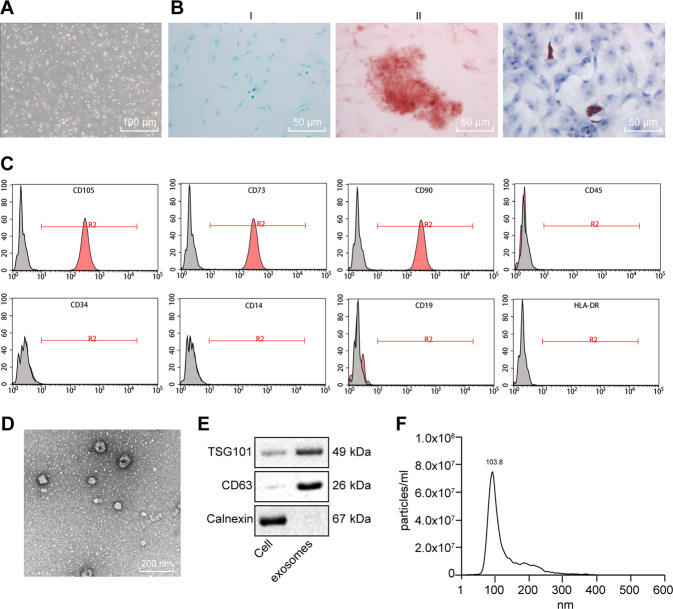
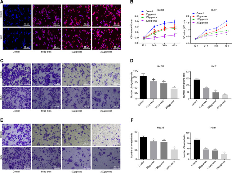
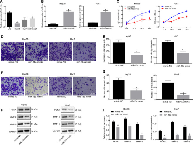
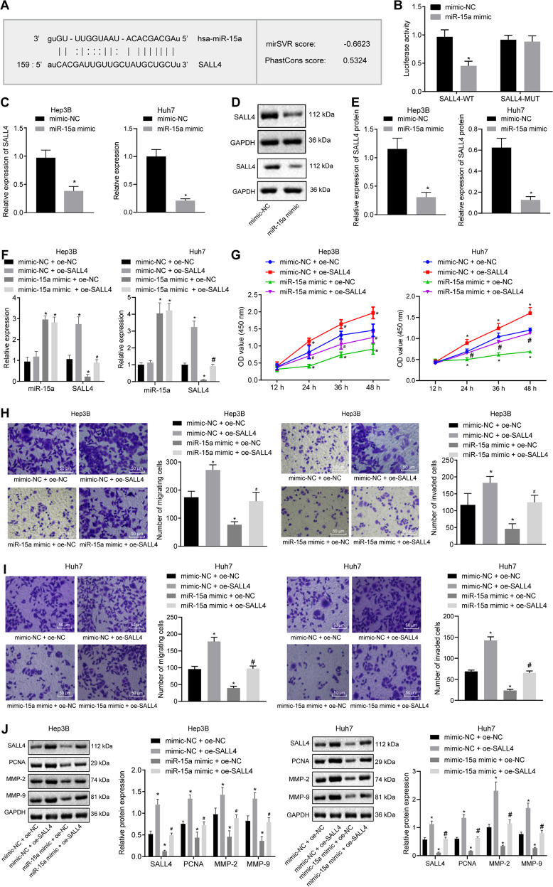
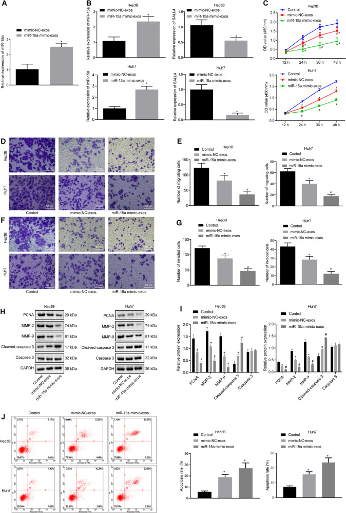
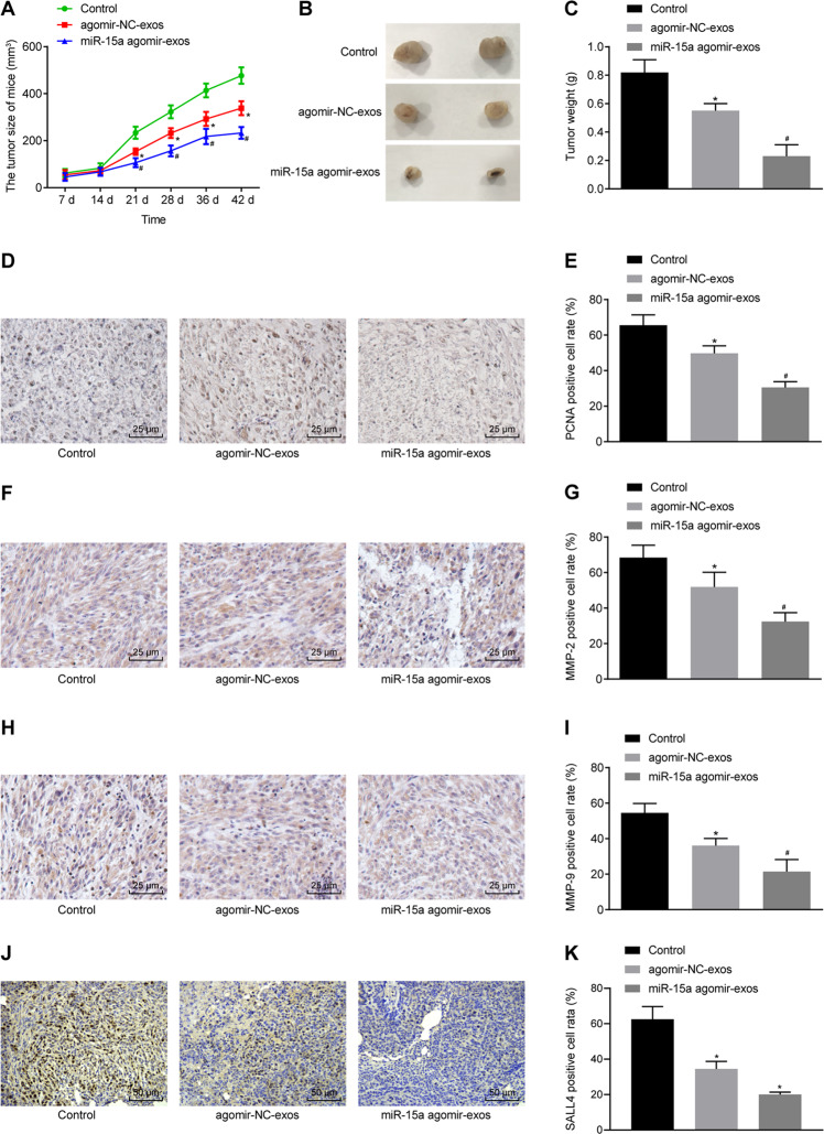
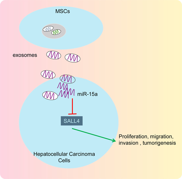
Hepatocellular carcinoma (HCC) is a heterogeneous tumor with an increased incidence worldwide accompanied by high mortality and dismal prognosis. Emerging evidence indicates that mesenchymal stem cells (MSCs)-derived exosomes possess protective effects against various human diseases by transporting microRNAs (miRNAs or miRs). We aimed to explore the role of exosomal miR-15a derived from MSCs and its related mechanisms in HCC. Exosomes were isolated from transduced MSCs and co-incubated with Hep3B and Huh7 cells. miR-15a expression was examined by RT-qPCR in HCC cells, MSCs, and secreted exosomes. CCK-8, transwell, and flow cytometry were used to detect the effects of miR-15a or spalt-like transcription factor 4 (SALL4) on cell proliferative, migrating, invasive, and apoptotic properties. A dual-luciferase reporter gene assay was performed to validate the predicted targeting relationship of miR-15a with SALL4. Finally, in vivo experiments in nude mice were implemented to assess the impact of exosome-delivered miR-15a on HCC. The exosomes from MSCs restrained HCC cell proliferative, migrating, and invasive potentials, and accelerated their apoptosis. miR-15a was expressed at low levels in HCC cells and could bind to SALL4, thus curtailing the proliferative, migrating, and invasive abilities of HCC cells. Exosomes successfully delivered miR-15a to HCC cells. Exosomal miR-15a depressed tumorigenicity and metastasis of HCC tumors in vivo. Overall, exosomal miR-15a from MSCs can downregulate SALL4 expression and thereby retard HCC development.


Conflict of interest statement

The authors declare no competing interests.

Figures

Fig. 1
Fig. 1. Identification of MSCs and exosomes.
A Morphologic characteristics of MSCs at the fifth in culture were observed under an inverted microscope (×100). B Chondrocyte formation, osteoblast formation, and adipogenic differentiation were analyzed by cytochemical staining of Alcian blue (I, ×200), Alizarin Red, (II, ×200) and oil red O (III, ×200). C Expression of the surface marker proteins associated with MSCs was determined by flow cytometry. D Ultrastructure of exosomes was observed under a transmission electron microscope (×5000). E Representative western blots of the surface marker TSG101 and CD63 proteins. F Concentration and size of exosomes were detected by NTA analysis.
Fig. 2
Fig. 2. MSC-derived exosomes repress proliferation, migration, and invasion of HCC cells in vitro.
A Uptake of MSC-derived exosomes from Hep3B and Huh7 cells was observed under a laser confocal microscope (×200). B Effect of exosomes on the proliferation of Hep3B and Huh7 cells was detected by CCK-8 assay. C Effect of exosomes on the migration of Hep3B cells and Huh7 was detected by transwell assay (×200). D Statistical analysis of panel C. E Effect of exosomes on the invasion of Hep3B and Huh7 cells was detected by transwell assay (×200). F Statistical analysis of panel E. p < 0.05 vs. control cells. Data in panel B were analyzed by repeated-measures ANOVA with Bonferroni post hoc testing, and in panels, D and F were analyzed by one-way ANOVA followed by Tukey’s post hoc test. Data are shown as mean ± standard deviation of three technical replicates.
Fig. 3
Fig. 3. miR-15a suppresses the proliferation, migration, and invasion of HCC cells in vitro.
A Expression of miR-15a was determined by RT-qPCR in normal hepatocytes L-02 and HCC cells Hep3B, Huh7, and SMMC-7721, relative to U6. B Expression of miR-15a was determined by RT-qPCR in Hep3B and Huh7 cells upon miR-15a mimic transfection, relative to U6. C Proliferation of Hep3B and Huh7 cells was detected by CCK-8 assay upon miR-15a mimic transfection. D Migration of Hep3B and Huh7 cells was detected by transwell assay upon miR-15a mimic transfection (×200). E Statistical analysis of panel D. F Invasion of Hep3B and Huh7 cells was detected by transwell assay upon miR-15a mimic transfection (×200). G Statistical analysis of panel F. H Representative western blots of MMP-2, MMP-9, and PCNA proteins in miR-15a mimic-transfected Hep3B and Huh7 cells. I The quantitation of panel H. *p < 0.05 vs. L-02 cells or Hep3B and Huh7 cells transfected with mimic-NC. Data in panel C were analyzed by repeated-measures ANOVA with Bonferroni post hoc testing, data in panels B, E, G and I were analyzed by unpaired t-test, and data in panel A were assessed by one-way ANOVA followed by Tukey’s post hoc test. Data are shown as mean ± standard deviation of three technical replicates.
Fig. 4
Fig. 4. miR-15a reduces proliferation, migration, and invasion of HCC cells via SALL4 inhibition in vitro.
A Putative miR-15a binding sites in the 3’UTR of SALL4 mRNA in the miRDB website (http://www.mirdb.org/). B miR-15a binding to SALL4 in cells verified by dual luciferase reporter gene assay. C mRNA expression of SALL4 was determined by RT-qPCR in miR-15a mimic-transfected Hep3B and Huh7 cells, relative to GAPDH. D Representative western blots of SALL4 protein in miR-15a mimic-transfected Hep3B and Huh7 cells, relative to GAPDH. E The quantitation of panel D. F Expression of miR-15a and SALL4 was determined by RT-qPCR in Hep3B and Huh7 cells transfected with miR-15a mimic and/or on-SALL4, relative to U6. G Proliferation of Hep3B and Huh7 cells were detected by CCK-8 following transfection with miR-15a mimic and/or oe-SALL4. H Migration of Hep3B and Huh7 cells was detected by transwell assay following transfection with miR-15a mimic and/or oe-SALL4 (×200). I Invasion of Hep3B and Huh7 cells was detected by transwell assay following transfection with miR-15a mimic and/or oe-SALL4 (×200). J Representative western blots of SALL4, MMP-2, MMP-9 and PCNA proteins in Hep3B and Huh7 cells following transfection with miR-15a mimic and/or oe-SALL4, relative to GAPDH. *p < 0.05 vs. Hep3B and Huh7 cells transfected with mimic-NC; #p < 0.05 vs. Hep3B and Huh7 cells transfected with miR-15a mimic+ of-NC. Data in panel G were analyzed by repeated-measures ANOVA with Bonferroni post hoc test. and data in panels BE were analyzed by unpaired t-test. Data in panels F, H, I, and J were analyzed by one-way ANOVA followed by Tukey’s post hoc test. Data are shown as mean ± standard deviation of three technical replicates.
Fig. 5
Fig. 5. Exosomes transfer miR-15a to HCC cells and thus affect proliferation, migration, invasion, and apoptosis of HCC cells.
A Expression of miR-15a was determined by RT-qPCR in exosomes from the miR-15a mimic-treated MSCs, relative to U6 (*p < 0.05 vs. exosomes from the MSCs transfected with mimic-NC). B Expression of miR-15a and SALL4 was determined by RT-qPCR in the co-culture system of Hep3B and Huh7 cells and miR-15a mimic-exos, relative to U6 and GAPDH, respectively. C Hep3B and Huh7 cell proliferation was detected by CCK-8 following co-culture with miR-15a mimic-exos. D Hep3B, and Huh7 cell migration were detected by Transwell assay following co-culture with miR-15a mimic-exos (×200). E Statistical analysis of panel D. F Hep3B and Huh7 cell invasion was detected by Transwell assay following co-culture with miR-15a mimic-exos (×200). G Statistical analysis of panel F. H Representative western blots of Cle-caspase3, caspase3, MMP-2, MMP-9, and PCNA proteins in the co-culture system of Hep3B and Huh7 cells and miR-15a mimic-exos, relative to GAPDH. I The quantitation of panel H. J The apoptosis of Hep3B and Huh7 cells detected using flow cytometry. *p < 0.05 vs. control Hep3B and Huh7 cells; #p < 0.05 vs. Hep3B and Huh7 cells co-cultured with mimic-NC-exos. Data in panel C were analyzed by repeated-measures ANOVA with Bonferroni post hoc test, and data in panels A and B were analyzed by unpaired t-test. Data in panels E, G, I, and J were analyzed by one-way ANOVA, with Tukey’s post-hoc test. Data are shown as mean ± standard deviation of three technical replicates.
Fig. 6
Fig. 6. Delivery of miR-15a by exosomes prevents tumor growth in vivo.
A The growth of the HCC xenograft tumor was measured every 7 days in nude mice injected with Hep3B cells treated with miR-15a agomir-exos. B Macroscopic observation of xenograft tumor of nude mice injected with Hep3B cells treated with miR-15a agomir-exos. C Tumor weight of nude mice injected with Hep3B cells treated with miR-15a agomir-exos. D Positive expression rate of PCNA protein was examined by immunohistochemistry in tumor tissues of nude mice injected with Hep3B cells treated with miR-15a agomir-exos (×400). E The quantitation of panel D. F Positive expression rate of MMP-2 protein was examined by immunohistochemistry in tumor tissues of nude mice injected with Hep3B cells treated with miR-15a agomir-exos (×400). G The quantitation of panel F. H Positive expression rate of MMP-9 protein was examined by immunohistochemistry in tumor tissues of nude mice injected with Hep3B cells treated with miR-15a agomir-exos (×400). I The quantitation of panel H. J Positive expression rate of SALL4 protein was examined by immunohistochemistry in tumor tissues of nude mice injected with Hep3B cells treated with miR-15a agomir-exos (×400). K The quantitation of panel J. *p < 0.05 vs. control mice; #p < 0.05 vs. mice injected with agomir-NC-exos-treated Hep3B cells. Data in panel A were analyzed by repeated-measures ANOVA with Bonferroni test, and data in panels C, E, G, I, and K were analyzed by one-way ANOVA, with Tukey’s post-hoc test. n = 12 for mice following each treatment.
Fig. 7
Fig. 7. A schematic for the mechanism and function of MSC-derived exosomal miR-15a in HCC via SALL4.
MSC-derived exosomes were capable of transmitting miR-15a, which thus inhibited the expression of SALL4, and thereby inhibiting the proliferation, migration, and invasion of HCC cells in vitro as well as retarding the tumorigenesis in vivo, ultimately preventing the occurrence of HCC.

Similar articles

Cited by

References

    1. Sun X, Zhuo XB, Hu YP, Zheng X, Zhao QJ. A novel matrine derivative WM622 inhibits hepatocellular carcinoma by inhibiting PI3K/AKT signaling pathways. Mol Cell Biochem. 2018;449:47–54. doi: 10.1007/s11010-018-3341-9. - DOI - PubMed
    1. Beck M, Schirmacher P, Singer S. Alterations of the nuclear transport system in hepatocellular carcinoma—new basis for therapeutic strategies. J Hepatol. 2017;67:1051–61. doi: 10.1016/j.jhep.2017.06.021. - DOI - PubMed
    1. Yang JD, Hainaut P, Gores GJ, Amadou A, Plymoth A, Roberts LR. A global view of hepatocellular carcinoma: trends, risk, prevention and management. Nat Rev Gastroenterol Hepatol. 2019;16:589–604. doi: 10.1038/s41575-019-0186-y. - DOI - PMC - PubMed
    1. Fitzmorris P, Shoreibah M, Anand BS, Singal AK. Management of hepatocellular carcinoma. J Cancer Res Clin Oncol. 2015;141:861–76. doi: 10.1007/s00432-014-1806-0. - DOI - PMC - PubMed
    1. Yang C, Yang H, Liu J, Zhu L, Yu S, Zhang X, et al. Focus on exosomes: novel pathogenic components of leukemia. Am J Cancer Res. 2019;9:1815–29. - PMC - PubMed