Review
. 2021 Aug 28;32(9):115.
doi: 10.1007/s10856-021-06583-x.

Biomimetic mineralisation systems for in situ enamel restoration inspired by amelogenesis

Affiliations
Review

Biomimetic mineralisation systems for in situ enamel restoration inspired by amelogenesis

Jue Wang et al. J Mater Sci Mater Med. .

Abstract

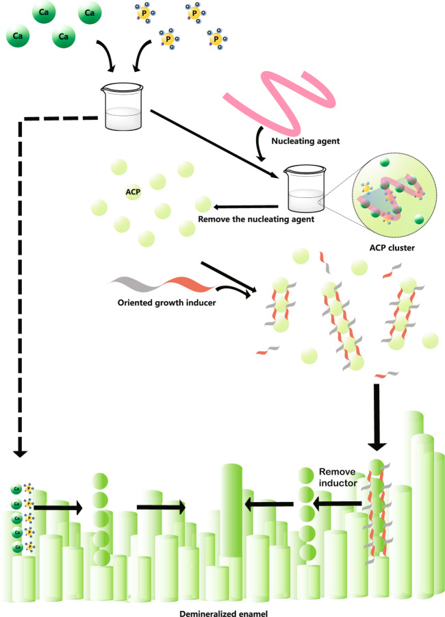
Caries and dental erosion are common oral diseases. Traditional treatments involve the mechanical removal of decay and filling but these methods are not suitable for cases involving large-scale enamel erosion, such as hypoplasia. To develop a noninvasive treatment, promoting remineralisation in the early stage of caries is of considerable clinical significance. Therefore, biomimetic mineralisation is an ideal approach for restoring enamel. Biomimetic mineralisation forms a new mineral layer that is tightly attached to the surface of the enamel. This review details the state-of-art achievements on the application of amelogenin and non-amelogenin, amorphous calcium phosphate, ions flow and other techniques in the biomimetic mineralisation of enamel. The ultimate goal of this review was to shed light on the requirements for enamel biomineralisation. Hence, herein, we summarise two strategies of biological minimisation systems for in situ enamel restoration inspired by amelogenesis that have been developed in recent years and compare their advantages and disadvantages.


Conflict of interest statement

The authors declare no competing interests.

Figures

Fig. 1
Fig. 1
A schematic model of the formation of ACP and transform into enamel-like crystals for biomineralisation. Strategy A (solid arrow): CaP will be stabilised by nucleating agent and form into ACP clusters. After the removement of the agent, the ACP will transform into crystals. Strategy B (dashed arrow): flow of CaP are selected or accelerated into the deep lesions, then nucleate to form ACP and finally transform into HAP
Fig. 2
Fig. 2
Enamel matrix subunit compartments from native enamel matrix and in ameloblast secretory vesicles imaged using aberration-corrected scanning transmission electron microscopy (STEM) imaging. A and B are STEM images of different parts of the natural enamel of 1 day postnatal developing mouse molars. Crystal nucleation in the compartment of organic subunits in natural enamel matrix can be seen in both A and B. Note the individual 1–3 nm diameter nucleation sites (arrows, A, B) randomly dispersed with a circular 20 nm diameter organic matrix assembly (matrix) (A, B).
Fig. 3
Fig. 3
Microstructure of enamel-like apatite crystals in the newly grown layer. Scanning electron microscopy (SEM) images showing A, B etched enamel, newly grown hydroxyapatite crystals in CS (C, D), AMEL-CS hydrogel (E, F) and AMEL-CS-MMP-20 (G, H). Arrows in D, H and F indicate the crystal orientation [33]
Fig. 4
Fig. 4
Transmission electron microscopy (TEM) images of nanospheres formed from peptides P26 (d) and P32 (e) at pH 7.4 at 25 °C. Reproduced with permission from ref. [40]
Fig. 5
Fig. 5
SEM images of the restored HAP layers treated in P26 after 7 days of incubation in artificial saliva in pH 7.0 at 37 °C. a Cross-sectional view of regenerated HAP layers. b Magnified image of (a) (yellow square) depicts the newly formed perpendicularly stacked crystals with a seamless attachment interface with underlying enamel rods. Reproduced with permission from ref. [40]
Fig. 6
Fig. 6
Restoration of the complicated structure of enamel by CMC-ALN/ACP-Gly. SEM results showing the surface morphology of normal enamel in (a). The surface morphologies of demineralized enamel (b, c); remineralised enamel with CMC/ACP nanocomplexes (d, e); remineralised enamel with CMC-ALN/ACP nanocomplexes (f, g); remineralised enamel with combination of CMC-ALN/ACP nanocomplexes and Gly (h–k), showing layers of oriented and ordered mineral crystals perpendicular to the enamel surface indicated with arrows of white dash line, meanwhile, on these layers scattered mineral crystals parallel on the enamel surface indicated with arrows of red dash line. Reproduced with permission from ref. [16]
Fig. 7
Fig. 7
Restoration of the complicated structure of enamel by TEA. A SEM image showing both etched enamel and repaired enamel. B A three-dimensional atomic force microscopy (AFM) image of repaired enamel. C High-magnification SEM image of the red circle in (A). D Cross-sectional view of final repaired enamel, where both enamel rods and inter-rods were repaired. Rods and IR represent for enamel rod and inter-rod, respectively. E, F Enamel rods with different orientations can be repaired [7]
Fig. 8
Fig. 8
Schematic of the ADGS. The PDA-modified substrates were coated by AAO membrane, a calcium-containing gelatin hydrogel and an ion-free gelatin hydrogel, from bottom to top. Phosphate solution was added to the top for mineralisation through ion diffusion. Reproduced with permission from ref. [73]
Fig. 9
Fig. 9
Three-dimensional X-ray mercury cadmium telluride images of an EAER-treated lesion. EAER-treated lesion reducing in volume and size. The red colouring represents the area of the enamel lesion that had a calculated mineral density ≥5% lower than the surrounding ‘healthy’ enamel [82]
Fig. 10
Fig. 10
SEM examinations of demineralised enamel and EAER-treated enamel. In the untreated lesions, the enamel rod structure is clear: rods are broken and demineralized. However, enamel in the EAER-treated lesions is very similar to nature enamel, with no degraded rods or broken rods visible [82]

Similar articles

Cited by

References

    1. Selwitz RH, Ismail AI, Pitts NB. Dental caries. Lancet. 2007;369:51–9. doi: 10.1016/s0140-6736(07)60031-2. - DOI - PubMed
    1. La Fontaine A, Zavgorodniy A, Liu H, Zheng R, Swain M, Cairney J. Atomic-scale compositional mapping reveals Mg-rich amorphous calcium phosphate in human dental enamel. Sci Adv. 2016;2:e1601145. doi: 10.1126/sciadv.1601145. - DOI - PMC - PubMed
    1. Moradian-Oldak J. Protein-mediated enamel mineralization. Front Biosci (Landmark Ed) 2012;17:1996–2023. doi: 10.2741/4034. - DOI - PMC - PubMed
    1. Palmer LC, Newcomb CJ, Kaltz SR, Spoerke ED, Stupp SI. Biomimetic systems for hydroxyapatite mineralization inspired by bone and enamel. Chem Rev. 2008;108:4754–83. doi: 10.1021/cr8004422. - DOI - PMC - PubMed
    1. Onuma K, Yamagishi K, Oyane A. Nucleation and growth of hydroxyapatite nanocrystals for nondestructive repair of early caries lesions. J Cryst Growth. 2005;282:199–207. doi: 10.1016/j.jcrysgro.2005.04.085. - DOI

MeSH terms

LinkOut - more resources