

Multiclass classification of whole-body scintigraphic images using a self-defined convolutional neural network with attention modules
- PMID: 34455613
- PMCID: PMC9135133
- DOI: 10.1002/mp.15196
Multiclass classification of whole-body scintigraphic images using a self-defined convolutional neural network with attention modules
Abstract
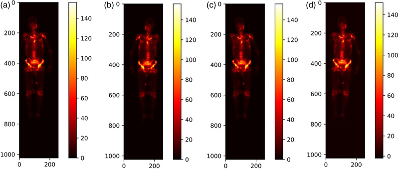
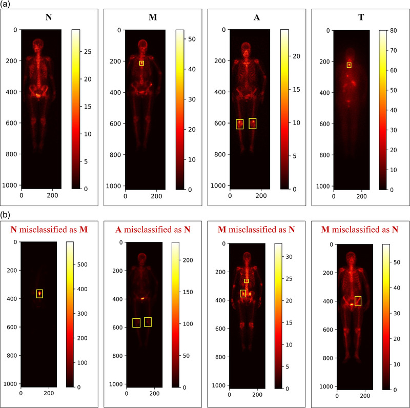
Purpose: A self-defined convolutional neural network is developed to automatically classify whole-body scintigraphic images of concern (i.e., the normal, metastasis, arthritis, and thyroid carcinoma), automatically detecting diseases with whole-body bone scintigraphy.
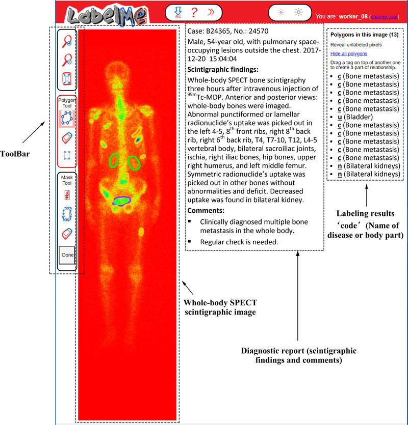
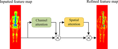
Methods: A set of parameter transformation operations are first used to augment the original dataset of whole-body bone scintigraphic images. A hybrid attention mechanism including the spatial and channel attention module is then introduced to develop a deep classification network, Dscint, which consists of eight weight layers, one hybrid attention module, two normalization modules, two fully connected layers, and one softmax layer.
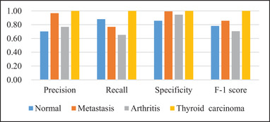
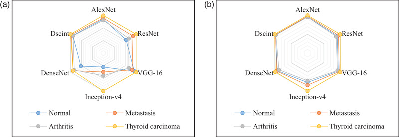
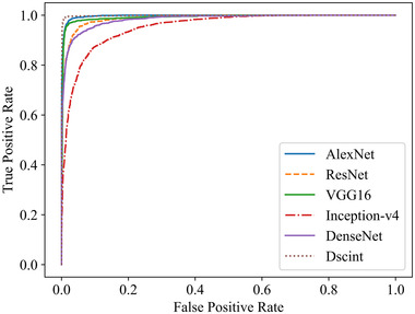
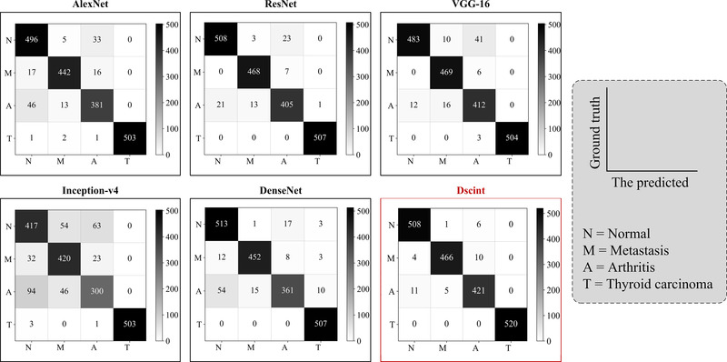
Results: Experimental evaluations conducted on a set of whole-body scintigraphic images show that the proposed deep classification network, Dscint, performs well for automated detection of diseases by classifying the images of concerns, achieving the accuracy, precision, recall, specificity, and F-1 score of 0.9801, 0.9795, 0.9791, 0.9933, and 0.9792, respectively, on the test data in the augmented dataset. A comparative analysis of Dscint and several classical deep classification networks (i.e., AlexNet, ResNet, VGGNet, DenseNet, and Inception-v4) reveals that our self-defined network, Dscint, performs best on classifying whole-body scintigraphic images on the same dataset.
Conclusions: The self-defined deep classification network, Dscint, can be utilized to automatically determine whether a whole-body scintigraphic image is either normal or contains diseases of concern. Specifically, better performance of Dscint is obtained on images with lesions that are present in relatively fixed locations like thyroid carcinoma than those with lesions occurring in nonfixed locations of bone tissue.
Keywords: attention mechanism; bone scintigraphy; convolutional neural network; medical image analysis; multiclass classification.
© 2021 American Association of Physicists in Medicine.
Conflict of interest statement
The authors declare no conflict of interest.
Figures

References
-
- Lin Q, Man Z, Cao Y, et al. Classifying functional nuclear images with convolutional neural networks: a survey. IET Image Proc. 2020;14(14):3300‐3313.
-
- Pi Y, Zhao Z, Xiang Y, et al. Automated diagnosis of bone metastasis based on multi‐view bone scans using attention‐augmented deep neural networks. Med Image Anal. 2020;65:101784. - PubMed
MeSH terms
Grants and funding
- 2021QB-063/Youth Ph.D. Foundation of Education Department of Gansu Province
- 31920210013/Fundamental Research Funds for the Central Universities
- 20JR5RA511/Natural Science Foundation of Gansu Province
- 61562075/National Natural Science Foundation of China
- 11080305/Gansu Provincial First-class Discipline Program of Northwest Minzu University
