Review
. 2021 Dec;12(1):90-114.
doi: 10.1080/19491034.2021.1962610.

Nuclear envelope mechanobiology: linking the nuclear structure and function

Affiliations
Review

Nuclear envelope mechanobiology: linking the nuclear structure and function

Matthew Goelzer et al. Nucleus. 2021 Dec.

Abstract

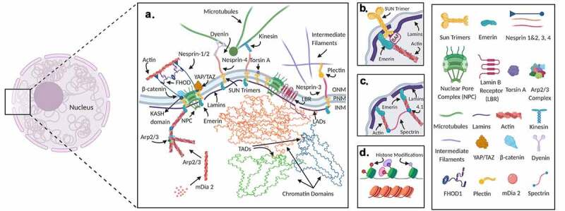
The nucleus, central to cellular activity, relies on both direct mechanical input as well as its molecular transducers to sense external stimuli and respond by regulating intra-nuclear chromatin organization that determines cell function and fate. In mesenchymal stem cells of musculoskeletal tissues, changes in nuclear structures are emerging as a key modulator of their differentiation and proliferation programs. In this review we will first introduce the structural elements of the nucleoskeleton and discuss the current literature on how nuclear structure and signaling are altered in relation to environmental and tissue level mechanical cues. We will focus on state-of-the-art techniques to apply mechanical force and methods to measure nuclear mechanics in conjunction with DNA, RNA, and protein visualization in living cells. Ultimately, combining real-time nuclear deformations and chromatin dynamics can be a powerful tool to study mechanisms of how forces affect the dynamics of genome function.

Keywords: Nuclear envelope; chromatin; live imaging; mechanobiology; nuclear mechanics.


Conflict of interest statement

No potential conflict of interest was reported by the authors.

Figures

Figure 1.
Figure 1.
Nucleus is a mechanically integrated mechanosignaling center. Nuclear structural proteins interact with the cytoskeleton, chromatin, and the nuclear membrane to stabilize the nucleus and provide mechanosensing functions (Insert A). LINC complexes composed of Sun 1/2 trimers and Nesprin 1/2 mechanically couple the actin cytoskeleton. The LINC complex also interacts with nuclear pore complexes (NPC) and in-part regulate the access of important mechanical transducers such as β-catenin and YAP/TAZ into the nucleus. Nesprin-3 through interactions with plectin and nesprin-4 are also known to interact with cytoplasmic intermediate filaments and microtubules, respectively. Nesprins can also bind to microtubules via dynein and kinesin. Mechanical coupling of actin and the LINC complex involves cytoplasmic formins such as FHOD1 that attaches nesprins and actin at multiple points for a more robust association. Torsin A may also facilitate the LINC assembly at the nuclear envelope. A nuclear envelope transmembrane protein, Emerin connects the LINC complex, via SUN1/2 and nesprin-1/2 to the chromatin through BAF and lamin A/C (Insert B). Emerin also associates and plays a role in regulating extra and intranuclear actin. The intranuclear actin network is formed through the crosslinking of short F-actin fibers via protein 4.1 and spectrin that provides elastic structural properties to the nucleus (Insert C). Inside the nucleus, G-actin is assembled into linear and branched networks through regulatory proteins such as arp2/3 and mDia2 and influence chromatin dynamics and gene access. Chromatin domains that bind to the nuclear lamins are called lamin-associated-domains (LAD). These domains have been shown to be correlated with heterochromatin, producing repression of gene expression of genes in the LADs. These chromatin domains conserve epigenetic histone modifications. Changes of histone modifications, topologically associated domains (TADs), and LADs all result in changes in gene expression and cell differentiation (Insert D)

Similar articles

Cited by

References

    1. Kanchanawong P, Shtengel G, Pasapera AM, et al. Nanoscale architecture of integrin-based cell adhesions. Nature. 2010;468(7323):580–584. - PMC - PubMed
    1. Burridge K, Fath K, Kelly T, et al. Focal adhesions: transmembrane junctions between the extracellular matrix and the cytoskeleton. Annu Rev Cell Biol. 1988;4(1):487–525. - PubMed
    1. Grashoff C, Hoffman BD, Brenner MD, et al. Measuring mechanical tension across vinculin reveals regulation of focal adhesion dynamics. Nature. 2010;466(7303):263–266. - PMC - PubMed
    1. Turner CE, Glenney JR, Burridge K.. Paxillin: a new vinculin-binding protein present in focal adhesions. J Cell Biol. 1990;111(3):1059–1068. - PMC - PubMed
    1. Pasapera AM, Schneider IC, Rericha E, et al. Myosin II activity regulates vinculin recruitment to focal adhesions through FAK-mediated paxillin phosphorylation. J Cell Biol. 2010;188(6):877–890. - PMC - PubMed

Publication types

LinkOut - more resources