. 2020 Dec:45:1-45.
doi: 10.3767/persoonia.2020.45.01. Epub 2019 Jun 18.

Cytospora (Diaporthales) in China

Affiliations

Cytospora (Diaporthales) in China

X L Fan et al. Persoonia. 2020 Dec.

Abstract

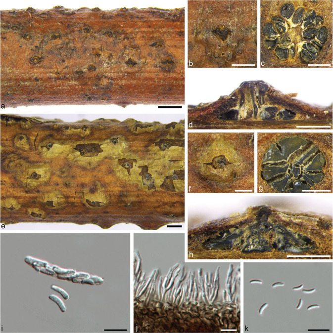
Members of the genus Cytospora are often reported as endophytes, saprobes or phytopathogens, primarily causing canker diseases of woody host plants. They occur on a wide range of hosts and have a worldwide distribution. Although several species have in the past been reported from China, the vast majority are not known from culture or DNA phylogeny. The primary aim of the present study was thus to clarify the taxonomy and phylogeny of a large collection of Cytospora species associated with diverse hosts in China. Cytospora spp. were collected in northeast, northwest, north and southwest China, indicating that the cold and dry environments favour these fungi. In this paper, we provide an assessment of 52 Cytospora spp. in China, focussing on 40 species represented by 88 isolates from 28 host genera. Based on a combination of morphology and a six-locus phylogeny (ITS, LSU, act1, rpb2, tef1-α and tub2), 13 new species and one new combination are introduced. The majority of the species investigated here appear to be host-specific, although further collections and pathogenicity studies will be required to confirm this conclusion.

Keywords: Valsa; canker disease; new taxa; plant pathogen; systematics; taxonomy.


Figures

Fig. 1
Fig. 1
Disease symptoms associated with Cytospora spp. a–b. Populus alba subsp. pyramidalis; c. Ailanthus altissima; d. Euonymus maackii; e–f. Juglans regia.
Fig. 2
Fig. 2
Infection process of Cytospora chrysosperma on Populus alba subsp. pyramidalis. a. Uninfected tissues; b. initial stage of pycnidial development (aggregation of mycelial cells in the cortex tissues) (6 d); c. formation of locules in the centre of pycnidial primordium (10 d); d–e. combination of adjacent locules (12 d); f. mycelial mass above locules rupturing the cuticle and forming an ostiole through the apical rupture (15 d). — Scale bars = 0.5 mm.
Fig. 3
Fig. 3
Maximum parsimony phylogram of Cytospora obtained from the ITS rDNA gene matrix. MP bootstrap support values above 50 % and posterior probabilities above 0.95 from BI are given at the first and second position. — Scale bar = 20 nucleotide substitutions. Ex-type strains are in bold; strains obtained in the current study in blue.
Fig. 3
Fig. 3
Maximum parsimony phylogram of Cytospora obtained from the ITS rDNA gene matrix. MP bootstrap support values above 50 % and posterior probabilities above 0.95 from BI are given at the first and second position. — Scale bar = 20 nucleotide substitutions. Ex-type strains are in bold; strains obtained in the current study in blue.
Fig. 4
Fig. 4
Maximum parsimony phylogram of Cytospora obtained from the combined matrix of ITS, LSU, act1, rpb2, tef1-α and tub2 genes. MP bootstrap support values above 50 % and posterior probabilities above 0.95 from BI are given at the first and second position. — Scale bar = 200 nucleotide substitutions. Ex-type strains are in bold; strains obtained in the current study in blue.
Fig. 4
Fig. 4
Maximum parsimony phylogram of Cytospora obtained from the combined matrix of ITS, LSU, act1, rpb2, tef1-α and tub2 genes. MP bootstrap support values above 50 % and posterior probabilities above 0.95 from BI are given at the first and second position. — Scale bar = 200 nucleotide substitutions. Ex-type strains are in bold; strains obtained in the current study in blue.
Fig. 5
Fig. 5
Cytospora ailanthicola on Ailanthus altissima (BJFC-S550). a–c. Habit of conidiomata on branch; d–e. transverse section through conidiomata; f–g. longitudinal section through conidiomata; h–i. conidiogenous cells with attached conidia; j. conidia. — Scale bars: a = 1 mm; b–g = 0.5 mm; h–j = 10 μm.
Fig. 6
Fig. 6
Cytospora atrocirrhata on Juglans regia (BJFC-S649). a–b. Habit of conidiomata on branch; c. transverse section through conidioma; d. longitudinal section through conidioma; e. conidiogenous cells with attached conidia; f. conidia. — Scale bars: a = 1 mm; b–d = 0.5 mm; e–f = 10 μm.
Fig. 7
Fig. 7
Cytospora beilinensis on Pinus armandii (BJFC-S1108). a. Disease symptoms; b–c. habit of conidiomata on branch; d. transverse section through conidioma; e. longitudinal section through conidioma; f. conidiogenous cells with attached conidia; g. conidia. — Scale bars: a = 5 mm; c–e = 0.5 mm; f–g = 10 μm.
Fig. 8
Fig. 8
Cytospora berberidis on Berberis dasystachya (BJFC-S681). a–b. Habit of conidiomata on branch; c. transverse section through conidioma; d. longitudinal section through conidioma; e. conidiogenous cells with attached conidia; f. conidia. — Scale bars: a = 1 mm; b–d = 0.5 mm; e–f = 10 μm.
Fig. 9
Fig. 9
Cytospora bungeanae on Pinus bungeana (BJFC-S1106). a–b. Habit of conidiomata on branch; c. transverse section through conidioma; d. longitudinal section through conidioma; e. conidiogenous cells with attached conidia; f. conidia. — Scale bars: a = 1 mm; b–d = 0.5 mm; e–f = 10 μm.
Fig. 10
Fig. 10
Cytospora carbonacea on Ulmus pumila (BJFC-S630). a–b. Habit of ascostromata on branch; c. transverse section through ascostroma; d. longitudinal section through ascostroma; e–f. habit of conidiomata on branch; g. transverse section through conidioma; h. longitudinal section through conidioma; i–j. asci; k. ascospores; l. conidiogenous cells with attached conidia; m. conidia. — Scale bars: a, e = 1 mm; b–d, f–h = 0.5 mm; i–m = 10 μm.
Fig. 11
Fig. 11
Cytospora celtidicola on Celtis sinensis (BJFC-S1107). a–b. Habit of conidiomata on branch; c. transverse section through conidioma; d. longitudinal section through conidioma; e. conidiogenous cells with attached conidia; f. conidia. — Scale bars: a = 1 mm; b–d = 0.5 mm; e–f = 10 μm.
Fig. 12
Fig. 12
Cytospora ceratosperma on Juglans regia (BJFC-S774). a–b. Habit of conidiomata on branch; c. transverse section through conidioma; d. longitudinal section through conidioma; e. conidiogenous cells with attached conidia; f. conidia. — Scale bars: a = 1 mm; b–d = 0.5 mm; e–f = 10 μm.
Fig. 13
Fig. 13
Cytospora ceratospermopsis on Juglans regia (BJFC-S567). a–b. Habit of conidiomata on branch; c. transverse section through conidioma; d. longitudinal section through conidioma; e. conidiogenous cells with attached conidia; f. conidia. — Scale bars: a = 1 mm; b–d = 0.5 mm; e–f = 10 μm.
Fig. 14
Fig. 14
Cytospora chrysosperma on Populus alba subsp. pyramidalis (BJFC-S750). a–b. Habit of ascostromata on branch; c. transverse section through ascostroma; d. longitudinal section through ascostroma; e–f. habit of conidiomata on branch; g. transverse section through conidioma; h. longitudinal section through conidioma; i. ascus; j. ascospores; k–l. conidiogenous cells with attached conidia; m. conidia. — Scale bars: a, e = 1 mm; b–d, f–h = 0.5 mm; i–m = 10 μm.
Fig. 15
Fig. 15
Cytospora elaeagni on Elaeagnus angustifolia (BJFC-S965). a–b. Habit of conidiomata on branch; c. transverse section through conidioma; d. longitudinal section through conidioma; e. conidiogenous cells with attached conidia; f. conidia. — Scale bars: a = 1 mm; b–d = 0.5 mm; e–f = 10 μm.
Fig. 16
Fig. 16
Cytospora erumpens on Prunus padus (BJFC-S1064). a–b. Habit of ascostromata on branch; c. transverse section through ascostroma; d. longitudinal section through ascostroma; e–f. habit of conidiomata on branch; g. transverse section through conidioma; h. longitudinal section through conidioma; i. ascus; j. ascospores; k. conidiogenous cells with attached conidia; l. conidia. — Scale bars: a, e = 1 mm; b–d, f–h = 0.5 mm; i–l = 10 μm.
Fig. 17
Fig. 17
Cytospora euonymicola on Euonymus kiautschovicus (BJFC-S1105). a–b. Habit of conidiomata on branch; c. transverse section through conidioma; d. longitudinal section through conidioma; e. conidiogenous cells with attached conidia; f. conidia. — Scale bars: a = 1 mm; b–d = 0.5 mm; e–f = 10 μm.
Fig. 18
Fig. 18
Cytospora euonymina on Euonymus kiautschovicus (BJFC-S1025). a–b. Habit of conidiomata on branch; c. transverse section through conidioma; d. longitudinal section through conidioma; e. conidiogenous cells with attached conidia; f. conidia. — Scale bars: a = 1 mm; b–d = 0.5 mm; e–f = 10 μm.
Fig. 19
Fig. 19
Cytospora fortunea on Cryptomeria japonica (HSFC 15206). a, Herbarium material; b, d. habit of ascostromata on branch; c, g. habit of conidiomata on branch; e. transverse section through ascostroma; f. longitudinal section through ascostroma; h. transverse section through conidioma; i. longitudinal section through conidioma; j–k. asci; l. ascospores; m–n. conidia. — Scale bars: b–c = 1 mm; d–i = 0.5 mm; j–n = 10 μm.
Fig. 20
Fig. 20
Cytospora gigalocus on Juglans regia (BJFC-S647). a–b. Habit of conidiomata on branch; c. transverse section through conidioma; d. longitudinal section through conidioma; e. conidiogenous cells with attached conidia; f. conidia. — Scale bars: a = 1 mm; b–d = 0.5 mm; e–f = 10 μm.
Fig. 21
Fig. 21
Cytospora gigaspora on Salix psammophila (BJFC-S975). a–b. Habit of conidiomata on branch; c. transverse section through conidioma; d. longitudinal section through conidioma; e. conidiogenous cells with attached conidia; f. conidia. — Scale bars: a = 1 mm; b–d = 0.5 mm; e–f = 10 μm.
Fig. 22
Fig. 22
Cytospora hippophaës on Hippophaë rhamnoides (BJFC-S779). a–b. Habit of ascostromata on branch; c. transverse section through ascostroma; d. longitudinal section through ascostroma; e–f. habit of conidiomata on branch; g. transverse section through conidioma; h. longitudinal section through conidioma; i–j. asci; k. ascospores; l. conidiogenous cells with attached conidia; m. conidia. — Scale bars: a, e = 1 mm; b–d, f–h = 0.5 mm; i–m = 10 μm.
Fig. 23
Fig. 23
Cytospora japonica on Prunus cerasifera f. atropurpurea (BJFC-S832). a–b. Habit of conidiomata on branch; c. transverse section through conidioma; d. longitudinal section through conidioma; e. conidiogenous cells with attached conidia; f. conidia. — Scale bars: a = 1 mm; b–d = 0.5 mm; e–f = 10 μm.
Fig. 24
Fig. 24
Cytospora juniperina on Juniperus przewalskii (CF-2015410). a–b. Habit of ascostromata on branch; c. transverse section through ascostroma; d. longitudinal section through ascostroma; e–f. habit of conidiomata on branch; g. transverse section through conidioma; h. longitudinal section through conidioma; i–j. asci; k. ascospores; l. conidiogenous cells with attached conidia; m. conidia. — Scale bars: a, e = 1 mm; b–d, f–h = 0.5 mm; i–m = 10 μm.
Fig. 25
Fig. 25
Cytospora leucosperma on Pyrus sp. (BJFC-S618). a, b. Habit of conidiomata on branch; c. transverse section through conidioma; d. longitudinal section through conidioma; e. conidiogenous cells with attached conidia; f. conidia. — Scale bars: a = 1 mm; b–d = 0.5 mm; e–f = 10 μm.
Fig. 26
Fig. 26
Cytospora leucostoma on Prunus persica (BJFC-S918). a–b. Habit of ascostromata on branch; c. transverse section through ascostroma; d. longitudinal section through ascostroma; e–f. habit of conidiomata on branch; g. transverse section through conidioma; h. longitudinal section through conidioma; i. asci; j. ascospores; k. conidiogenous cells with attached conidia; l. conidia. — Scale bars: a, e = 1 mm; b–d, f–h = 0.5 mm; i–l = 10 μm.
Fig. 27
Fig. 27
Cytospora mali on Malus pumila (BJFC-S503). a–b. Habit of ascostromata on branch; c. transverse section through ascostroma; d. longitudinal section through ascostroma; e–f. habit of conidiomata on branch; g. transverse section through conidioma; h. longitudinal section through conidioma; i–j. asci; k. ascospores; l. conidiogenous cells with attached conidia; m. conidia. — Scale bars: a, e = 1 mm; b–d, f–h = 0.5 mm; i–m = 10 μm.
Fig. 28
Fig. 28
Cytospora melnikii on Rhus typhina (BJFC-XJHJS). a. Host; b. habit of conidioma on branch; c. transverse section through conidioma; d. longitudinal section through conidioma; e–f. conidiogenous cells with attached conidia; g. conidia. — Scale bars: a = 1 mm; b–d = 0.5 mm; e–g = 10 μm.
Fig. 29
Fig. 29
Cytospora nivea on Populus alba subsp. pyramidalis (BJFC-S1307). a. Disease symptoms; b. habit of conidiomata on branch; c. transverse section through conidioma; d. longitudinal section through conidioma; e. conidiogenous cells with attached conidia; f. conidia. — Scale bars: a = 5 mm; b–d = 0.5 mm; e–f = 10 μm.
Fig. 30
Fig. 30
Cytospora platycladi on Platycladus orientalis (BJFC-S1111). a–b. Habit of conidiomata on branch; c. transverse section through conidioma; d. longitudinal section through conidioma; e. conidiogenous cells with attached conidia; f. conidia. — Scale bars: a = 1 mm; b–d = 0.5 mm; e–f = 10 μm.
Fig. 31
Fig. 31
Cytospora platycladicola on Platycladus orientalis (BJFC-S758). a–b. Habit of ascostromata on branch; c. transverse section through ascostroma; d. longitudinal section through ascostroma; e–f. habit of conidiomata on branch; g. transverse section through conidioma; h. longitudinal section through conidioma; i. asci; j. ascospores; k. conidiogenous cells with attached conidia; l. conidia. — Scale bars: a, e = 1 mm; b–d, f–h = 0.5 mm; i–l = 10 μm.
Fig. 32
Fig. 32
Cytospora populina on Salix psammophila (BJFC-S978). a–b. Habit of ascostromata on branch; c. transverse section through ascostroma; d. longitudinal section through ascostroma; e–g. asci (e, f immature); h. ascospores. — Scale bars: a = 1 mm; b–d = 0.5 mm; e–h = 10 μm.
Fig. 33
Fig. 33
Cytospora populinopsis on Sorbus aucuparia (BJFC-S802). a–b. Habit of ascostromata on branch; c. transverse section through ascostroma; d. longitudinal section through ascostroma; e–f. asci; g. ascospores. — Scale bars: a = 1 mm; b–d = 0.5 mm; e–g = 10 μm.
Fig. 34
Fig. 34
Cytospora pruinopsis on Ulmus pumila (BJFC-S1073). a–b. Habit of conidiomata on branch; c. transverse section through conidioma; d. longitudinal section through conidioma; e. conidiogenous cells with attached conidia; f. conidia. — Scale bars: a = 2 mm; b–d = 0.5 mm; e–f = 10 μm.
Fig. 35
Fig. 35
Cytospora pruinosa on Syringa oblata (BJFC-S636). a–b. Habit of conidiomata on branch; c. transverse section through conidioma; d. longitudinal section through conidioma; e. conidiogenous cells with attached conidia; f. conidia. — Scale bars: a = 1 mm; b–d = 0.25 mm; e–f = 10 μm.
Fig. 36
Fig. 36
Cytospora schulzeri on Malus pumila (BJFC-S538). a. Habit of conidiomata and ascostromata on branch; b. habit of ascostroma on branch; c. transverse section through ascostroma conidioma; d. longitudinal section through ascostroma; e. habit of conidioma on branch; f. transverse section through conidioma; g. longitudinal section through conidioma; h–i. asci; j. conidiogenous cells with attached conidia; k. conidia. — Scale bars: a = 1 mm; b–g = 0.5 mm; h–k = 10 μm.
Fig. 37
Fig. 37
Cytospora sibiraeae on Sibiraea angustata (BJFC-S783). a–b. Habit of ascostromata on branch; c. ectostromatic disc with ostioles; d. transverse section through ascostroma; e. longitudinal section through ascostroma; f–h. asci; i. ascospores. — Scale bars: a = 1 mm; b–e = 0.5 mm; f–i = 10 μm.
Fig. 38
Fig. 38
Cytospora sophorae on Styphnolobium japonicum (BJFC-S698). a–b. Habit of conidiomata on branch; c. transverse section through conidioma; d. longitudinal section through conidioma; e. conidiogenous cells with attached conidia; f. conidia. — Scale bars: a = 1 mm; b–d = 0.5 mm; e–f = 10 μm.
Fig. 39
Fig. 39
Cytospora sophoricola on Styphnolobium japonicum (BJFC-S694). a–b. Habit of conidiomata on branch; c. transverse section through conidioma; d. longitudinal section through conidioma; e. conidiogenous cells with attached conidia; f. conidia. — Scale bars: a = 1 mm; b–d = 0.5 mm; e–f = 10 μm.
Fig. 40
Fig. 40
Cytospora sophoriopsis on Styphnolobium japonicum (BJFC-S713). a–b. Habit of conidiomata on branch; c. transverse section through conidioma; d. longitudinal section through conidioma; e. conidiogenous cells with attached conidia; f. conidia. — Scale bars: a = 1 mm; b–d = 0.5 mm; e–f = 10 μm.
Fig. 41
Fig. 41
Cytospora tamaricicola on Tamarix chinensis (CF-2015510). a–b. Habit of ascostromata on branch; c. transverse section through ascostroma; d. longitudinal section through ascostroma; e–f. habit of conidiomata on branch; g. transverse section through conidioma; h. longitudinal section through conidioma; i. ascus and ascospores; j. conidiogenous cells with attached conidia; k. conidia. — Scale bars: a, e = 1 mm; b–d, f–h = 0.5 mm; i–k = 10 μm.

Similar articles

Cited by

References

    1. Adams GC, Roux J, Wingfield MJ, et al. . 2005. Phylogenetic relationships and morphology of Cytospora species and related teleomorphs (Ascomycota, Diaporthales, Valsaceae) from Eucalyptus. Studies in Mycology 52: 1–144.
    1. Adams GC, Roux J, Wingfield MJ. 2006. Cytospora species (Ascomycota, Diaporthales, Valsaceae), introduced and native pathogens of trees in South Africa. Australasian Plant Pathology 35: 521–548.
    1. Adams GC, Surve-Iyer RS, Iezzoni AF. 2002. Ribosomal DNA sequence divergence and group I introns within the Leucostoma species L. cinctum, L. persoonii, and L. parapersoonii sp. nov., ascomycetes that cause Cytospora canker of fruit trees. Mycologia 94: 947–967. - PubMed
    1. Ariyawansa HA, Hyde KD, Jayasiri SC, et al. . 2015. Fungal diversity notes 111–252 – taxonomic and phylogenetic contributions to fungal taxa. Fungal Diversity 75: 27–274.
    1. Barr ME. 1978. The Diaporthales in North America with emphasis on Gnomonia and its segregates. Mycologia Memoir 7: 1–232.

LinkOut - more resources