Review
. 2021 Aug 12:12:732742.
doi: 10.3389/fphys.2021.732742. eCollection 2021.

The Biogenesis Process of VDAC - From Early Cytosolic Events to Its Final Membrane Integration

Affiliations
Review

The Biogenesis Process of VDAC - From Early Cytosolic Events to Its Final Membrane Integration

Anasuya Moitra et al. Front Physiol. .

Abstract

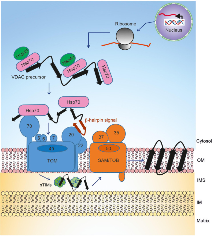
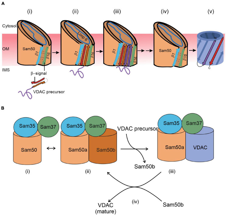
Voltage dependent anion-selective channel (VDAC) is the most abundant protein in the mitochondrial outer membrane. It is a membrane embedded β-barrel protein composed of 19 mostly anti-parallel β-strands that form a hydrophilic pore. Similar to the vast majority of mitochondrial proteins, VDAC is encoded by nuclear DNA, and synthesized on cytosolic ribosomes. The protein is then targeted to the mitochondria while being maintained in an import competent conformation by specific cytosolic factors. Recent studies, using yeast cells as a model system, have unearthed the long searched for mitochondrial targeting signal for VDAC and the role of cytosolic chaperones and mitochondrial import machineries in its proper biogenesis. In this review, we summarize our current knowledge regarding the early cytosolic stages of the biogenesis of VDAC molecules, the specific targeting of VDAC to the mitochondrial surface, and the subsequent integration of VDAC into the mitochondrial outer membrane by the TOM and TOB/SAM complexes.

Keywords: TOM complex; VDAC; beta-barrels; chaperones; mitochondria; outer membrane.


Conflict of interest statement

The authors declare that the research was conducted in the absence of any commercial or financial relationships that could be construed as a potential conflict of interest.

Figures

FIGURE 1
FIGURE 1
Biogenesis pathway of VDAC. Precursors of VDAC are transcribed in the nucleus, translated on cytosolic ribosomes, and then transported to the mitochondrial surface with the help of chaperones. At the outer membrane, the precursors are initially recognized by receptors of the TOM complex and then translocated across the membrane via the pore formed by Tom40. In the IMS, the small TIM chaperones relay the newly synthesized VDAC molecule to the TOB/SAM complex, which facilitates the final steps of membrane integration.
FIGURE 2
FIGURE 2
A working model for the final steps of the membrane integration of VDAC. (A) Lateral insertion (adapted from Figure 8; Höhr et al., 2018). (i) VDAC precursors approach the outer membrane from the IMS. (ii) The C-terminal β-signal of VDAC precursor interferes with the Sam50 structure by binding to the β1 strand of Sam50 and disrupting the β1–β16 interactions within Sam50. This enforces opening of a lateral gate. (iii) The initial opening is followed by sequential insertion of additional precursor β-hairpins through the lateral gate of Sam50. (iv) The lateral gate of Sam50 re-closes to (v) release the fully formed β-barrel of VDAC into the MOM. (B) Barrel switching (based on Takeda et al., 2021). In its substrate-free state, the SAM complex is in equilibrium between the “monomeric” state consisting of Sam50a, Sam35, and Sam37 (i) and a “dimeric” species that contains a second (Sam50b) barrel (ii). The gradually formed VDAC barrel displaces Sam50b (iii). Finally, the fully folded VDAC molecule dissociates from the complex to be replaced by Sam50b (iv).

References

    1. Araiso Y., Tsutsumi A., Qiu J., Imai K., Shiota T., Song J., et al. (2019). Structure of the mitochondrial import gate reveals distinct preprotein paths. Nature 575 395–401. 10.1038/s41586-019-1680-7 - DOI - PubMed
    1. Bausewein T., Mills D. J., Langer J. D., Nitschke B., Nussberger S., Kühlbrandt W. (2017). Cryo-EM structure of the TOM core complex from Neurospora crassa. Cell 170 693–700.e697. - PubMed
    1. Benz R. (1989). “Porins from mitochondrial and bacterial outer membranes: structural and functional aspects,” in Anion Carriers of Mitochondrial Membranes, eds Azzi A., Nałęz K. A., Nałęcz M. J., Wojtczak L. (Berlin: Springer; ).
    1. Beverly K. N., Sawaya M. R., Schmid E., Koehler C. M. (2008). The Tim8-Tim13 complex has multiple substrate binding sites and binds cooperatively to Tim23. J. Mol. Biol. 382 1144–1156. 10.1016/j.jmb.2008.07.069 - DOI - PMC - PubMed
    1. Colombini M. (1979). A candidate for the permeability pathway of the outer mitochondrial membrane. Nature 279 643–645. 10.1038/279643a0 - DOI - PubMed

LinkOut - more resources