

Adipocyte-Endothelium Crosstalk in Obesity
- PMID: 34456860
- PMCID: PMC8387580
- DOI: 10.3389/fendo.2021.681290
Adipocyte-Endothelium Crosstalk in Obesity
Abstract
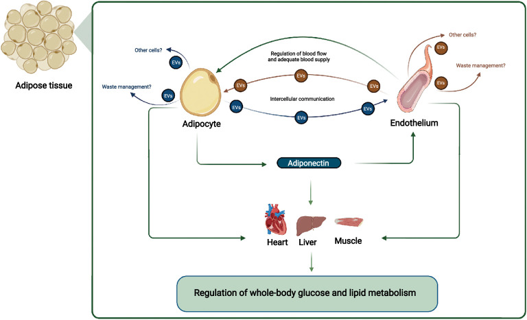
Obesity is characterized by pathological adipose tissue (AT) expansion. While healthy AT expansion enhances systemic insulin sensitivity, unhealthy AT expansion through increased adipocyte size is associated with insulin resistance, fibrosis, hypoxia, and reduced adipose-derived adiponectin secretion. The mechanisms causing the unhealthy AT expansion are not fully elucidated; yet, dysregulated crosstalk between cells within the AT is an important contributor. Evidence from animal and human studies suggests a crucial role of the crosstalk between vascular endothelium (the innermost cell type in blood vessels) and adipocytes for metabolic homeostasis. Arterial endothelial cells are directly involved in maintaining normal organ functions through local blood flow regulation. The endothelial-dependent regulation of blood flow in AT is hampered in obesity, which negatively affects the adipocyte. Moreover, endothelial cells secrete extracellular vesicles (EVs) that target adipocytes in vivo. The endothelial EVs secretion is hampered in obesity and may be affected by the adipocyte-derived adipokine adiponectin. Adiponectin targets the vascular endothelium, eliciting organ-protective functions through binding to T-cadherin. The reduced obesity-induced adiponectin binding of T-cadherin reduces endothelial EV secretion. This affects endothelial health and cell-cell communication between AT cells and distant organs, influencing systemic energy homeostasis. This review focuses on the current understanding of endothelial and adipocyte crosstalk. We will discuss how obesity changes the AT environment and how these changes contribute to obesity-associated metabolic disease in humans. Particularly, we will describe and discuss the EV-dependent communication and regulation between adipocytes, adiponectin, and the endothelial cells regulating systemic energy homeostasis in health and metabolic disease in humans.
Keywords: adipose tissue; endocrine; endothelial cells; extracellular vesicles; hypoxia; nitric oxide.
Copyright © 2021 Sabaratnam and Svenningsen.
Conflict of interest statement
The authors declare that the research was conducted in the absence of any commercial or financial relationships that could be construed as a potential conflict of interest.
Figures
Similar articles
-
Adipocyte-endothelial cell interplay in adipose tissue physiology.Biochem Pharmacol. 2024 Apr;222:116081. doi: 10.1016/j.bcp.2024.116081. Epub 2024 Feb 24. Biochem Pharmacol. 2024. PMID: 38408682 Review.
-
Increased secretion of adipocyte-derived extracellular vesicles is associated with adipose tissue inflammation and the mobilization of excess lipid in human obesity.J Transl Med. 2024 Jul 4;22(1):623. doi: 10.1186/s12967-024-05249-w. J Transl Med. 2024. PMID: 38965596 Free PMC article.
-
Role of perivascular adipose tissue in nicotine‑induced endothelial cell inflammatory responses.Mol Med Rep. 2016 Dec;14(6):5713-5718. doi: 10.3892/mmr.2016.5934. Epub 2016 Nov 9. Mol Med Rep. 2016. PMID: 27840948
-
Interplay between adipose tissue and blood vessels in obesity and vascular dysfunction.Rev Endocr Metab Disord. 2013 Mar;14(1):49-58. doi: 10.1007/s11154-012-9230-8. Rev Endocr Metab Disord. 2013. PMID: 23283583 Review.
-
Inflammatory adipocyte-derived extracellular vesicles promote leukocyte attachment to vascular endothelial cells.Atherosclerosis. 2019 Apr;283:19-27. doi: 10.1016/j.atherosclerosis.2019.01.013. Epub 2019 Jan 24. Atherosclerosis. 2019. PMID: 30771557
Cited by
-
Endotheliopathy in the metabolic syndrome: Mechanisms and clinical implications.Pharmacol Ther. 2023 Apr;244:108372. doi: 10.1016/j.pharmthera.2023.108372. Epub 2023 Mar 7. Pharmacol Ther. 2023. PMID: 36894027 Free PMC article. Review.
-
Zingiberaceae in Cardiovascular Health: A review of adipokine modulation and endothelial protection via adipocyte-endothelial crosstalk mechanism.Curr Nutr Rep. 2025 May 14;14(1):66. doi: 10.1007/s13668-025-00656-x. Curr Nutr Rep. 2025. PMID: 40366476 Review.
-
Obesity and hypertension: Obesity medicine association (OMA) clinical practice statement (CPS) 2023.Obes Pillars. 2023 Aug 7;8:100083. doi: 10.1016/j.obpill.2023.100083. eCollection 2023 Dec. Obes Pillars. 2023. PMID: 38125655 Free PMC article.
-
A Potential Interplay between HDLs and Adiponectin in Promoting Endothelial Dysfunction in Obesity.Biomedicines. 2022 Jun 7;10(6):1344. doi: 10.3390/biomedicines10061344. Biomedicines. 2022. PMID: 35740366 Free PMC article. Review.
-
Enhanced Angiogenesis in HUVECs Preconditioned with Media from Adipocytes Differentiated from Lipedema Adipose Stem Cells In Vitro.Int J Mol Sci. 2023 Sep 1;24(17):13572. doi: 10.3390/ijms241713572. Int J Mol Sci. 2023. PMID: 37686378 Free PMC article.
References
Publication types
MeSH terms
LinkOut - more resources
Full Text Sources
Medical