

A Negative (1,3)-β-D-Glucan Result Alone Is Not Sufficient to Rule Out a Diagnosis of Pneumocystis Pneumonia in Patients With Hematological Malignancies
- PMID: 34456893
- PMCID: PMC8386019
- DOI: 10.3389/fmicb.2021.713265
A Negative (1,3)-β-D-Glucan Result Alone Is Not Sufficient to Rule Out a Diagnosis of Pneumocystis Pneumonia in Patients With Hematological Malignancies
Abstract
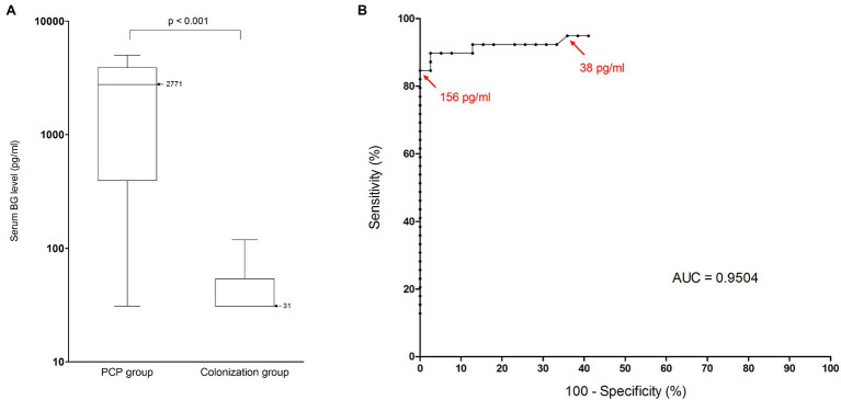
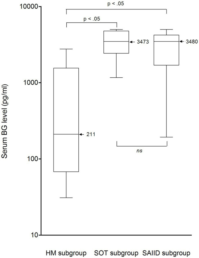
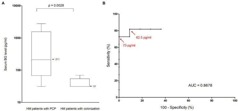
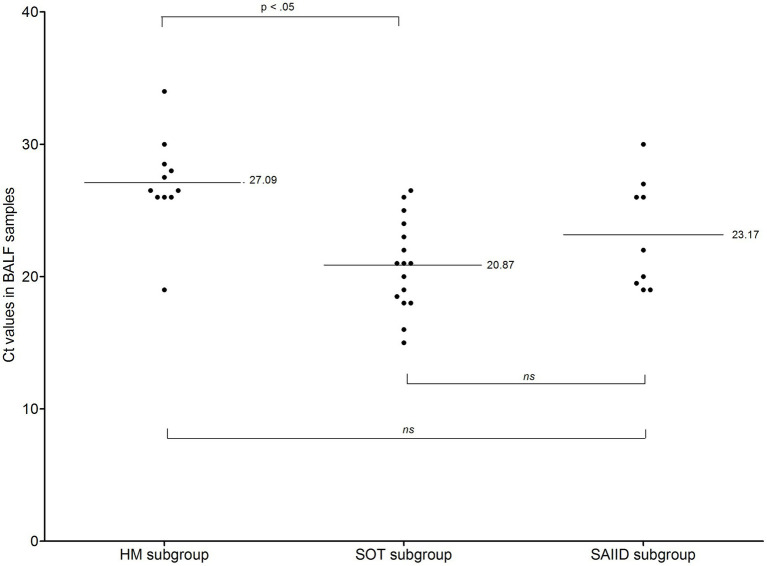
Background: Serum (1,3)-β-D-glucan (BG) testing is increasingly being used in the diagnostic armamentarium for invasive fungal diseases. Given its high sensitivity, some studies suggest that a negative BG result contributes to rule out a diagnosis of Pneumocystis pneumonia (PCP). However, recent reports described a suboptimal sensitivity in HIV-negative immunocompromised patients. In this study, we evaluated the performance of BG assay for PCP diagnosis in HIV-negative patients with diverse PCP risk factors. We also assessed the correlation between Pneumocystis jirovecii load in pulmonary samples and serum BG levels. Methods: We retrospectively included HIV-negative patients with microscopically proven PCP and for whom a BG result was available. We also enrolled patients colonized by Pneumocystis as control group. Colonized patients were matched with PCP patients based on their underlying condition that exposed to PCP. Pulmonary fungal loads were determined by an in-house real-time PCR, and BG levels were measured by using the Fungitell® kit (Associates of Cape Cod, Inc.). Results: Thirty-nine patients were included in each of the two groups. Thirty-four of 39 PCP patients and one of 39 colonized patient had a positive BG test, resulting in a sensitivity of 0.87 (95% CI: 0.73-0.94), a specificity of 0.97 (95% CI: 0.87-0.99), a positive predictive value of 0.97 (95% CI: 0.85-0.99), and a negative predictive value of 0.88 (95% CI: 0.75-0.95) for BG assay. Nonetheless, median BG level differed according to the underlying condition. Among the PCP group, the lowest median level of 211 pg/ml was observed in patients with hematological malignancy (HM) and differed significantly from that observed either in solid organ transplants (3,473 pg/ml) or in patients with autoimmune or inflammatory disorder (3,480 pg/ml). Indeed, the sensitivity of BG assay was estimated at 0.64 (95% CI: 0.35-0.85) in HM patients and was lower than the one observed in the whole PCP group. Furthermore, BG level and fungal burden correlated poorly among all PCP patients. Conclusion: BG is not a reliable biomarker for ruling out PCP in HIV-negative patients with HM. Interpretation of a negative BG result should take into account, but not be limited to, the underlying condition predisposing to PCP.
Keywords: (1,3)-β-D-glucan; HIV-negative patients; Pneumocystis jirovecii; Pneumocystis pneumonia; biomarker; bronchoalveolar lavage fluid; fungal load; hematological malignancies.
Copyright © 2021 Damiani, Demey, Pauc, Le Govic and Totet.
Conflict of interest statement
The authors declare that the research was conducted in the absence of any commercial or financial relationships that could be construed as a potential conflict of interest.
Figures
Similar articles
-
Variable reliability of the (1,3)-β-d-glucan test for screening Pneumocystis pneumonia in HIV-negative patients depending on the underlying condition.Med Mycol. 2024 Nov 12;62(11):myae106. doi: 10.1093/mmy/myae106. Med Mycol. 2024. PMID: 39504484
-
Combined quantification of pulmonary Pneumocystis jirovecii DNA and serum (1->3)-β-D-glucan for differential diagnosis of pneumocystis pneumonia and Pneumocystis colonization.J Clin Microbiol. 2013 Oct;51(10):3380-8. doi: 10.1128/JCM.01554-13. Epub 2013 Jul 31. J Clin Microbiol. 2013. PMID: 23903553 Free PMC article.
-
Usefulness of (1,3) ß-D-glucan detection in bronchoalveolar lavage samples in Pneumocystis pneumonia and Pneumocystis pulmonary colonization.J Mycol Med. 2015 Mar;25(1):36-43. doi: 10.1016/j.mycmed.2014.11.001. Epub 2014 Nov 7. J Mycol Med. 2015. PMID: 25498852
-
Molecular diagnosis of Pneumocystis pneumonia in immunocompromised patients.Curr Opin Infect Dis. 2019 Aug;32(4):314-321. doi: 10.1097/QCO.0000000000000559. Curr Opin Infect Dis. 2019. PMID: 31107250 Review.
-
Pneumocystis jirovecii pneumonia in non-HIV-infected patients in the era of novel immunosuppressive therapies.J Infect Chemother. 2012 Dec;18(6):793-806. doi: 10.1007/s10156-012-0453-0. Epub 2012 Aug 6. J Infect Chemother. 2012. PMID: 22864454 Review.
Cited by
-
Identifying optimal serum 1,3-β-D-Glucan cut-off for diagnosing Pneumocystis Jirovecii Pneumonia in non-HIV patients in the intensive care unit.BMC Infect Dis. 2024 Sep 20;24(1):1015. doi: 10.1186/s12879-024-09873-1. BMC Infect Dis. 2024. PMID: 39304817 Free PMC article.
-
Negative serum (1,3) -β-D-glucan has a low power to exclude Pneumocystis jirovecii pneumonia (PJP) in HIV-uninfected patients with positive qPCR.Ann Clin Microbiol Antimicrob. 2023 Nov 20;22(1):102. doi: 10.1186/s12941-023-00650-7. Ann Clin Microbiol Antimicrob. 2023. PMID: 37986091 Free PMC article.
-
An Evaluation of the OLM PneumID Real-Time Polymerase Chain Reaction to Aid in the Diagnosis of Pneumocystis Pneumonia.J Fungi (Basel). 2023 Nov 15;9(11):1106. doi: 10.3390/jof9111106. J Fungi (Basel). 2023. PMID: 37998911 Free PMC article.
-
Fungal β-Glucans: Biological Properties, Immunomodulatory Effects, Diagnostic and Therapeutic Applications.Infect Dis Clin Microbiol. 2025 Mar 27;7(1):1-16. doi: 10.36519/idcm.2025.448. eCollection 2025 Mar. Infect Dis Clin Microbiol. 2025. PMID: 40225707 Free PMC article. Review.
-
Beta-D-Glucan in Patients with Haematological Malignancies.J Fungi (Basel). 2021 Dec 7;7(12):1046. doi: 10.3390/jof7121046. J Fungi (Basel). 2021. PMID: 34947028 Free PMC article. Review.
References
-
- Alanio A., Hauser P. M., Lagrou K., Melchers W. J. G., Helweg-Larsen J., Matos O., et al. . (2016). ECIL guidelines for the diagnosis of Pneumocystis jirovecii pneumonia in patients with haematological malignancies and stem cell transplant recipients. J. Antimicrob. Chemother. 71, 2386–2396. 10.1093/jac/dkw156, PMID: - DOI - PubMed
-
- Damiani C., Le Gal S., Da Costa C., Virmaux M., Nevez G., Totet A. (2013). Combined quantification of pulmonary Pneumocystis jirovecii DNA and serum (1->3)-β-D-glucan for differential diagnosis of Pneumocystis pneumonia and Pneumocystis colonization. J. Clin. Microbiol. 51, 3380–3388. 10.1128/JCM.01554-13, PMID: - DOI - PMC - PubMed
-
- De Boer M. G. J., Gelinck L. B. S., van Zelst B. D., van de Sande W. W. J., Willems L. N. A., van Dissel J. T., et al. . (2011). β-D-glucan and S-adenosylmethionine serum levels for the diagnosis of Pneumocystis pneumonia in HIV-negative patients: a prospective study. J. Infect. 62, 93–100. 10.1016/j.jinf.2010.10.007, PMID: - DOI - PubMed
LinkOut - more resources
Full Text Sources