Review
. 2021 Aug 12:12:691766.
doi: 10.3389/fimmu.2021.691766. eCollection 2021.

The Functions of Hepatitis B Virus Encoding Proteins: Viral Persistence and Liver Pathogenesis

Affiliations
Review

The Functions of Hepatitis B Virus Encoding Proteins: Viral Persistence and Liver Pathogenesis

Fenglin Zhao et al. Front Immunol. .

Abstract

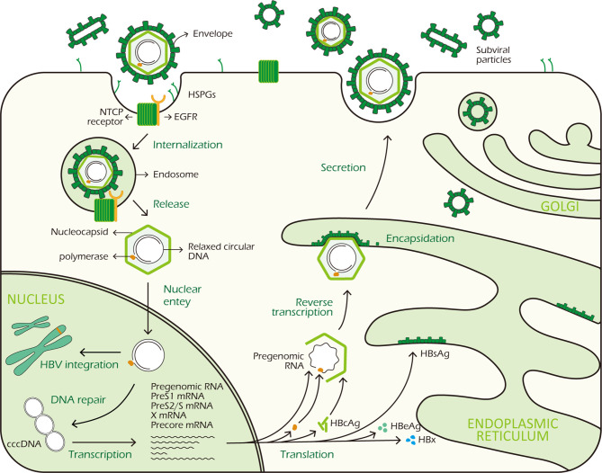
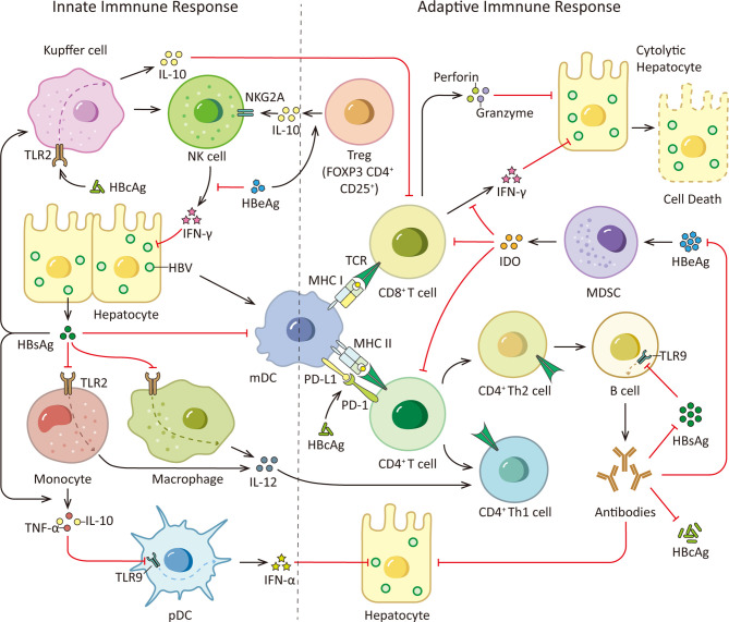
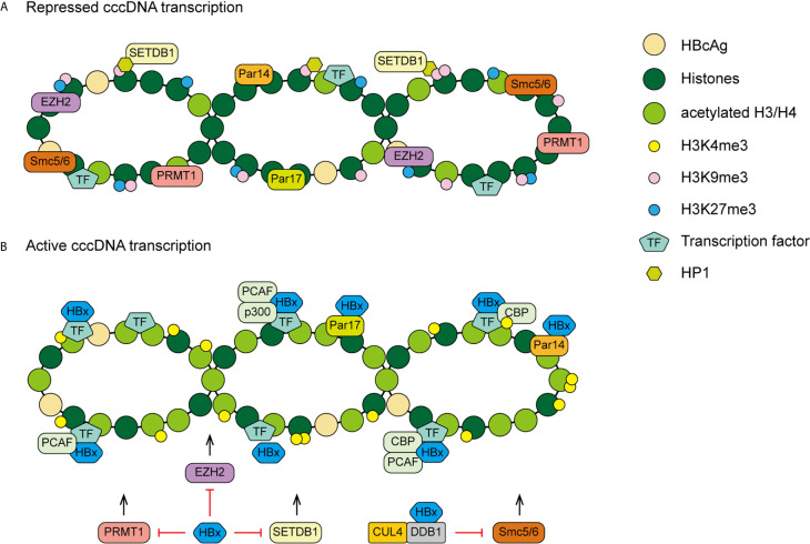
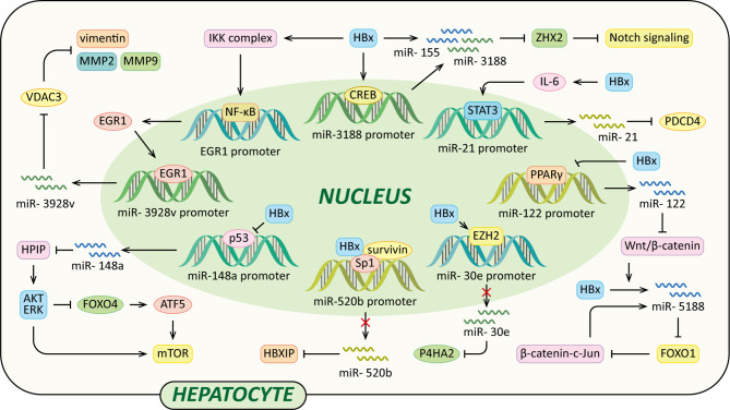
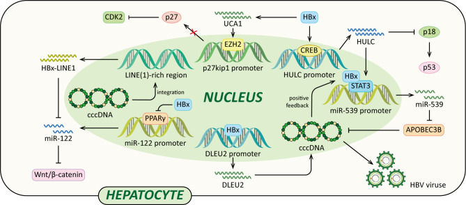
About 250 million people worldwide are chronically infected with Hepatitis B virus (HBV), contributing to a large burden on public health. Despite the existence of vaccines and antiviral drugs to prevent infection and suppress viral replication respectively, chronic hepatitis B (CHB) cure remains a remote treatment goal. The viral persistence caused by HBV is account for the chronic infection which increases the risk for developing liver cirrhosis and hepatocellular carcinoma (HCC). HBV virion utilizes various strategies to escape surveillance of host immune system therefore enhancing its replication, while the precise mechanisms involved remain elusive. Accumulating evidence suggests that the proteins encoded by HBV (hepatitis B surface antigen, hepatitis B core antigen, hepatitis B envelope antigen, HBx and polymerase) play an important role in viral persistence and liver pathogenesis. This review summarizes the major findings in functions of HBV encoding proteins, illustrating how these proteins affect hepatocytes and the immune system, which may open new venues for CHB therapies.

Keywords: encoding proteins; hepatitis B virus (HBV); hepatocellular carcinoma (HCC); immune response; viral escape.


Conflict of interest statement

The authors declare that the research was conducted in the absence of any commercial or financial relationships that could be construed as a potential conflict of interest.

Figures

Figure 1
Figure 1
HBV life cycle. HBV virus binds to HSPGs with low affinity to initiate infection, subsequently it interacts with NTCP receptor on the surface of hepatocyte to trigger viral internalization in a EGFR dependent manner. After being endocytosed, the virus releases its DNA-containing nucleocapsid into the hepatocyte cytoplasm which is subsequently transported to the nucleus. In the nucleus, the rcDNA is repaired and converted to cccDNA. At the same time, integration of viral DNA into host genome also takes place. The cccDNA minichromosome persists in the nucleus and functions as a transcription template for viral RNA transcription. It codes 7 viral proteins required for replication: 3 different sizes HBsAg, HBcAg, HBeAg, HBx and polymerase. In the cytoplasm, HBcAg proteins self-assemble into icosahedral nucleocapsid to package viral polymerase and pgRNA. After reverse transcription in nucleocapsid, HBV genome-containing capsid binds to HBsAg proteins in the ER for encapsidation. Finally, mature HBV viruses exit the hepatocytes via MVBs.
Figure 2
Figure 2
HBsAg, HBcAg, HBeAg and immune responses. During HBV infection, the viral PAMPs can be detected and recognized by innate immune cells, such as Kupffer cells, macrophages, monocytes and pDCs, contributing to the release of cytokines and the subsequent activation of other immune cells. However, HBV virus is able to counter this initial immune response with its proteome, especially HBsAg, HBeAg and HBcAg. HBsAg can selectively suppress the TLR2 ligand-stimulated IL-12 production in monocytes and macrophages, thus hinder IL-12 induced Th1 cell response and T cell proliferation. And it is able to induce monocytes to secrete TNF-α and IL-10 and down-regulate the TLR9 expression on pDCs, leading to decreased production of IFN-α by pDCs. Moreover, HBsAg inhibits up-regulation of costimulatory molecules during mDC maturation thus hinders mDC maturation and function. In addition, HBcAg can elevate IL-10 production via stimulating TLR2 on KCs which subsequently induce anti-HBV CD8+ T cell exhaustion. Viral clearance requires coordinated response of both the innate and adaptive immune system. Among adaptive immunity, vigorous T and B cell responses to the HBV encoding proteins is indispensable. While viral persistence is associated with impaired HBV-specific adaptive immune response. HBsAg decreases TLR9 transcription in B cells, resulting in diminished B cells proliferation, differentiation and antibodies production. Besides, HBcAg induces hyper-expression of PD-1 on CD4+ T cells and up-regulates the PD-L1 expression on monocyte-derived DC, leading to a coinhibitory signal to T cells. What’s more, HBeAg is able to induce elevated IL-10 production in Tregs, increasing expression of inhibitory receptor NKG2A on NK cells. And HBeAg induces mMDSCs expansion and up-regulates immune suppressor molecules IDO in mMDSCs to impair the proliferation of CD4+ and CD8+ T cells and IFN-γ production. TCR, T cell receptor.
Figure 3
Figure 3
A proposed model of HBx mediated cccDNA minichromosome epigenetic modulation. (A) In the absence of HBx, cccDNA-bound histones are hypoacetylated and the host restriction factors are recruited on the cccDNA, contributing to repressed gene transcription and viral replication. (B) In the presence of HBx, HATs are recruited to cccDNA and host restriction factors, such as PRMT1, EZH2, SETDB1 and Smc5/6, are inhibited by HBx, leading to hyperacetylation of cccDNA-bound histones and active cccDNA transcription.
Figure 4
Figure 4
Regulatory models of HBx and miRNAs involved in liver pathogenesis. The mechanisms involved in HBx and miRNAs are related to Notch signaling pathway, Wnt/β-catenin signaling pathway, MMP, collagen deposition, HPIP/AKT/ERK/FOXO4/ATF5/mTOR signaling pathway and HBXIP, which result in cell proliferation, anti-apoptosis, c-Jun activation, VM, EMT, liver fibrosis and eventually HCC.
Figure 5
Figure 5
Regulatory models of HBx and lncRNAs involved in liver pathogenesis. The mechanisms involved in HBx and lncRNAs are related to epigenetic modulation, Vimentin, miR-122, Wnt/β-catenin signaling pathway, p18, miR-539 and p27, which cause elevated HBV transcription and replication, EMT, diminished p53, cccDNA stability, cell growth, anti-apoptosis and eventually HCC.

Similar articles

Cited by

References

    1. World Health Organization . Global Hepatitis Report. (2017). Available at: https://www.who.int/publications/i/item/global-hepatitis-report-2017 (Accessed November 10, 2020).
    1. Tang LSY, Covert E, Wilson E, Kottilil S. Chronic Hepatitis B Infection: A Review. JAMA (2018) 319(17):1802–13. 10.1001/jama.2018.3795 - DOI - PubMed
    1. Roth GA, Abate D, Abate KH, Abay SM, Abbafati C, Abbasi N, et al. . Global, Regional, and National Age-Sex-Specific Mortality for 282 Causes of Death in 195 Countries and Territories, 1980–2017: A Systematic Analysis for the Global Burden of Disease Study 2017. Lancet (2018) 392(10159):1736–88. 10.1016/s0140-6736(18)32203-7 - DOI - PMC - PubMed
    1. Nassal M. HBV cccDNA: Viral Persistence Reservoir and Key Obstacle for a Cure of Chronic Hepatitis B. Gut (2015) 64(12):1972–84. 10.1136/gutjnl-2015-309809 - DOI - PubMed
    1. Tsai KN, Kuo CF, Ou JJ. Mechanisms of Hepatitis B Virus Persistence. Trends Microbiol (2018) 26(1):33–42. 10.1016/j.tim.2017.07.006 - DOI - PMC - PubMed

Publication types

LinkOut - more resources