. 2021 Aug 11:12:715704.
doi: 10.3389/fimmu.2021.715704. eCollection 2021.

Different Aspects of Classical Pathway Overactivation in Patients With C3 Glomerulopathy and Immune Complex-Mediated Membranoproliferative Glomerulonephritis

Affiliations

Different Aspects of Classical Pathway Overactivation in Patients With C3 Glomerulopathy and Immune Complex-Mediated Membranoproliferative Glomerulonephritis

Marloes A H M Michels et al. Front Immunol. .

Abstract

The rare and heterogeneous kidney disorder C3 glomerulopathy (C3G) is characterized by dysregulation of the alternative pathway (AP) of the complement system. C3G is often associated with autoantibodies stabilizing the AP C3 convertase named C3 nephritic factors (C3NeF). The role of classical pathway (CP) convertase stabilization in C3G and related diseases such as immune complex-mediated membranoproliferative glomerulonephritis (IC-MPGN) remains largely unknown. Here, we investigated the CP convertase activity in patients with C3G and IC-MPGN. Using a refined two-step hemolytic assay, we measured the stability of CP convertases directly in the serum of 52 patients and 17 healthy controls. In four patients, CP convertase activity was prolonged compared to healthy controls, i.e. the enzymatic complex was stabilized. In three patients (2 C3G, 1 IC-MPGN) the convertase stabilization was caused by immunoglobulins, indicating the presence of autoantibodies named C4 nephritic factors (C4NeFs). Importantly, the assay also enabled detection of non-immunoglobulin-mediated stabilization of the CP convertase in one patient with C3G. Prolonged CP convertase activity coincided with C3NeF activity in all patients and for up to 70 months of observation. Crucially, experiments with C3-depleted serum showed that C4NeFs stabilized the CP C3 convertase (C4bC2a), that does not contain C3NeF epitopes. All patients with prolonged CP convertase activity showed clear signs of complement activation, i.e. lowered C3 and C5 levels and elevated levels of C3d, C3bc, C3bBbP, and C5b-9. In conclusion, this work provides new insights into the diverse aspects and (non-)immunoglobulin nature of factors causing CP convertase overactivity in C3G/IC-MPGN.

Keywords: C3 convertase; C3 glomerulopathy; C3 nephritic factors (C3NeFs); C4 nephritic factors (C4NeFs); autoantibody; classical complement pathway.


Conflict of interest statement

JW has received grants from Achillion and Chemocentryx, outside the submitted work. The remaining authors declare that the research was conducted in the absence of any commercial or financial relationships that could be construed as a potential conflict of interest.

Figures

Figure 1
Figure 1
Flow chart of sample inclusion. All patient samples were measured for prolonged classical pathway activity, i.e. C4 nephritic factor (C4NeF) activity, but only the 52 patients with a final diagnosis of C3 glomerulopathy (C3G) or immune complex-mediated membranoproliferative glomerulonephritis (IC-MPGN) were included in this study. C3 nephritic factor (C3NeF) activity, i.e. prolonged alternative pathway activity, was measured in the number of patients indicated between brackets. DDD, dense deposit disease; C3GN, C3 glomerulonephritis; MGRS, monoclonal gammopathy of renal significance; HUS, hemolytic uremic syndrome; aHUS, atypical HUS; SLE, systemic lupus erythematosus.
Figure 2
Figure 2
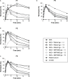
Classical pathway convertase activity in healthy controls. Classical pathway convertase activity was measured by incubating sensitized sheep erythrocytes with sera obtained from 17 healthy individuals. Normal human serum (NHS) and heat-inactivated NHS (hi-NHS) were used as assay controls. Hemolysis levels are presented as percentage of full lysis of erythrocytes in water. Results represent means of at least three independent measurements for each sample. Standard deviations have been omitted for better visibility.
Figure 3
Figure 3
Prolonged classical pathway convertase activity profiles in four patients. Classical pathway convertase activity was measured by incubating sensitized sheep erythrocytes with normal human serum (NHS) or with patient samples. Depicted are the activity profiles for patients P1, P2, P3, and P4, that tested positive for prolonged convertase activity as their top/t10 ratios were <1.6 in at least three measurements performed. Representative runs of these samples are shown. Data for NHS are given as the mean and range from the four experiments in which the positive patients were detected. The area under the NHS curve is indicated in grey. Hemolysis levels are presented as percentage of full lysis of erythrocytes in water.
Figure 4
Figure 4
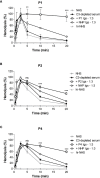
Classical pathway convertase activity profiles of control serum supplemented with patient immunoglobulin fractions. Sensitized sheep erythrocytes were incubated with normal human serum (NHS) mixed with an equal (1:1) or excess volume (1:3 and 1:5) of immunoglobulins (Igs) purified from the EDTA-plasma of patient P1 (A) and P4 (C) or from the serum of P2 (B) and P3 (D). As a negative control, Igs purified from normal human plasma (NHP) or NHS, depending on the source of the patient Igs, were added in similar ratios. The patient sera were used as positive controls. Hemolysis levels are presented as percentage of full lysis of erythrocytes in water. Concentration series were tested once. hi-NHS, heat-inactivated NHS.
Figure 5
Figure 5
Classical pathway C3 convertase (C4b2a) stabilization by patient immunoglobulins. Sensitized sheep erythrocytes were incubated with C3-depleted serum supplemented with immunoglobulins (Igs) from P1 (A), P2 (B), or P4 (C) in a 1:3 volume ratio. Igs from normal human plasma (NHP) were used as a control. Hemolysis levels are presented as percentage of full lysis of erythrocytes in water. Data are presented as the mean and the standard deviation of three independent experiments. Significance values according to two-way analysis of variance with Bonferroni post-test with C3-depleted serum + NHP Igs as the control condition are given for t5, t10, and t20: ***P < 0.001; **P < 0.01; ns, not significant. NHS, normal human serum; hi-NHS, heat-inactivated NHS.
Figure 6
Figure 6
C4 nephritic factor and C3 nephritic factor activity of patient P1 during follow-up. Samples from P1 collected over the disease course were tested for prolonged classical pathway activity, i.e. C4 nephritic factor (C4NeF) activity, and for prolonged alternative pathway activity, i.e. C3 nephritic factor (C3NeF) activity. Collection dates are presented as time after acute presentation in months (+mo). Hemolysis levels are presented as percentage of full lysis of erythrocytes in water. Results are shown as mean and range of two independent experiments. For the C4NeF activity assay, the positive control (PC) is the sample of P1 collected 32 mo after presentation. Part Rem, partial remission; NHS, normal human serum; hi-NHS, heat-inactivated NHS.
Figure 7
Figure 7
C4 nephritic factor and C3 nephritic factor activity of patients P2, P3, and P4 during follow-up. Samples from P2 (A), P3 (B), and P4 (C) collected over the disease course were tested for prolonged classical pathway activity, i.e. C4 nephritic factor (C4NeF) activity, and for prolonged alternative pathway activity, i.e. C3 nephritic factor (C3NeF) activity. Collection dates are presented as time after acute presentation in months (+mo). Hemolysis levels are presented as percentage of full lysis of erythrocytes in water. Results are shown as mean and range of two independent experiments. Part Rem, partial remission; PC, positive control; NHS, normal human serum; hi-NHS, heat-inactivated NHS.
Figure 8
Figure 8
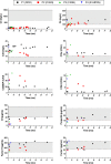
Complement activation profiles of the patients with prolonged classical pathway convertase activity during follow-up. Follow-up is indicated as time after initial presentation in months (mo). Grey areas indicate the healthy control ranges. DDD, dense deposit disease; C3GN, C3 glomerulonephritis; IC-MPGN, immune complex-mediated membranoproliferative glomerulonephritis; CAU, complement arbitrary units.

References

    1. Pickering MC, D’Agati VD, Nester CM, Smith RJ, Haas M, Appel GB, et al. . C3 Glomerulopathy: Consensus Report. Kidney Int (2013) 84(6):1079–89. 10.1038/ki.2013.377 - DOI - PMC - PubMed
    1. Smith RJH, Appel GB, Blom AM, Cook HT, D’Agati VD, Fakhouri F, et al. . C3 Glomerulopathy - Understanding a Rare Complement-Driven Renal Disease. Nat Rev Nephrol (2019) 15(3):129–43. 10.1038/s41581-018-0107-2 - DOI - PMC - PubMed
    1. Goodship TH, Cook HT, Fakhouri F, Fervenza FC, Fremeaux-Bacchi V, Kavanagh D, et al. . Atypical Hemolytic Uremic Syndrome and C3 Glomerulopathy: Conclusions From a “Kidney Disease: Improving Global Outcomes” (KDIGO) Controversies Conference. Kidney Int (2017) 91(3):539–51. 10.1016/j.kint.2016.10.005 - DOI - PubMed
    1. Fakhouri F, Fremeaux-Bacchi V, Noel LH, Cook HT, Pickering MC. C3 Glomerulopathy: A New Classification. Nat Rev Nephrol (2010) 6(8):494–9. 10.1038/nrneph.2010.85 - DOI - PubMed
    1. Servais A, Noël LH, Roumenina LT, Le Quintrec M, Ngo S, Dragon-Durey MA, et al. . Acquired and Genetic Complement Abnormalities Play a Critical Role in Dense Deposit Disease and Other C3 Glomerulopathies. Kidney Int (2012) 82(4):454–64. 10.1038/ki.2012.63 - DOI - PubMed

Publication types

MeSH terms