. 2021 Aug 11:12:709736.
doi: 10.3389/fgene.2021.709736. eCollection 2021.

Correction of Vertebral Bone Development in Ectodysplasin A1-Deficient Mice by Prenatal Treatment With a Replacement Protein

Affiliations

Correction of Vertebral Bone Development in Ectodysplasin A1-Deficient Mice by Prenatal Treatment With a Replacement Protein

Clara-Sophie Kossel et al. Front Genet. .

Abstract

X-linked hypohidrotic ectodermal dysplasia with the cardinal symptoms hypodontia, hypotrichosis and hypohidrosis is caused by a genetic deficiency of ectodysplasin A1 (EDA1). Prenatal EDA1 replacement can rescue the development of skin appendages and teeth. Tabby mice, a natural animal model of EDA1 deficiency, additionally feature a striking kink of the tail, the cause of which has remained unclear. We studied the origin of this phenomenon and its response to prenatal therapy. Alterations in the distal spine could be noticed soon after birth, and kinks were present in all Tabby mice by the age of 4 months. Although their vertebral bones frequently had a disorganized epiphyseal zone possibly predisposing to fractures, cortical bone density was only reduced in vertebrae of older Tabby mice and even increased in their tibiae. Different availability of osteoclasts in the spine, which may affect bone density, was ruled out by osteoclast staining. The absence of hair follicles, a well-known niche of epidermal stem cells, and much lower bromodeoxyuridine uptake in the tail skin of 9-day-old Tabby mice rather suggest the kink being due to a skin proliferation defect that prevents the skin from growing as fast as the skeleton, so that caudal vertebrae may be squeezed and bent by a lack of skin. Early postnatal treatment with EDA1 leading to delayed hair follicle formation attenuated the kink, but did not prevent it. Tabby mice born after prenatal administration of EDA1, however, showed normal tail skin proliferation, no signs of kinking and, interestingly, a normalized vertebral bone density. Thus, our data prove the causal relationship between EDA1 deficiency and kinky tails and indicate that hair follicles are required for murine tail skin to grow fast enough. Disturbed bone development appears to be partially pre-determined in utero and can be counteracted by timely EDA1 replacement, pointing to a role of EDA1 also in osteogenesis.

Keywords: NF-κB; bone; development; ectodermal dysplasia; ectodysplasin A1; fetal therapy.


Conflict of interest statement

PS, CK-Q, and HS are inventors on patents relevant to this publication. The remaining authors declare that the research was conducted in the absence of any commercial or financial relationships that could be construed as a potential conflict of interest.

Figures

FIGURE 1
FIGURE 1
Tabby mice develop vertebral dislocations soon after birth. Macroscopic comparison of representative tail tips from (A) wild-type and (B) untreated Tabby mice at embryonic day E18.5 and postnatal days P0, P4, and P21. Lower panels in panels (A,B) depict whole-mount staining of cartilaginous parts of the same tails with alcian blue. In nearly half of the Tabby mice, first alterations of the distal spine could be detected already on day P4; kinks were evident in 21 of 23 animals investigated at day P21 and in all Tabby mice by day P120.
FIGURE 2
FIGURE 2
Impact of ectodysplasin A1 (EDA1) deficiency on osteogenesis and bone density. (A,B) Representative three-dimensional reconstructions of (A) the distal tail portion and (B) tibiae of wild-type and Tabby mice. (C–F) Vertebral and tibial bone sections stained with hematoxylin-eosin (C,D) or alcian blue (E,F): The epiphyseal region appears less well organized and contains more cartilage islands (arrowed) in the tails of Tabby mice than in wild-type mice. (G) Although computed tomography scanning (micro-CT) revealed a tendency toward reduced cortical bone density already in tails of Tabby mice at an age of 2–4 months (mean of 7–10 distal vertebrae where the kink is formed; indicated in Hounsfield units), this difference became significant only in Tabby mice older than 6 months (n = 6; white bars) compared with wild-type animals (n = 6; black bars). (H) Tibial bones, in contrast, showed a higher cortical density in Tabby mice of both age groups (n = 6 and n = 7, respectively; white bars) than in wild-type animals (black bars) as determined by micro-CT measurements. Scale bar in panels (C–F): 100 μm. Data are shown as mean ± SD; p < 0.05; ****p < 0.0001.
FIGURE 3
FIGURE 3
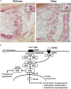
Possible role of EDA1 in the differentiation of osteoclasts. (A) Representative tartrate-resistant acid phosphatase (TRAP)-stained tail sections of wild-type and Tabby mice indicating similar number and distribution of osteoclasts in the metaphyseal trabecular part of vertebral bones. The boxes in the upper right corner show magnified osteoclasts (dark red). (B) Overview of the molecular link between the EDA1 pathway and the RANK–TRAF6–NFκB pathway and its possible involvement in osteoclastic differentiation. Scale bar: 100 μm.
FIGURE 4
FIGURE 4
Diminished proliferation of tail skin in Tabby mice. (A) Immunofluorescence staining of tail tip cross-sections from wild-type (n = 6; left panel) and Tabby mice (n = 6; right panel) after intraperitoneal injection of bromodeoxyuridine (BrdU) at day P9 to label replicating cells. The amount of BrdU-positive cells in the skin (red; nuclei counter-stained blue with DAPI) was assessed. (B,C) Quantitation of replicating cells visualized by BrdU incorporation. BrdU-positive nuclei were counted in the epidermis and the upper dermis (depth ≤ 0.15 mm) except for the hair bulb region. Data from six independent skin sections of each Tabby and wild-type mouse are shown. Scale bar: 50 μm. ****p < 0.0001.
FIGURE 5
FIGURE 5
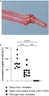
Attenuation of the tail kink in Tabby mice upon postnatal treatment with EDA1. (A) Tabby mice were treated at birth with EDA1 (2 mg/kg body weight, intraperitoneal injection) and sacrificed at weaning. Pictures of the tails were taken for measurements. The amplitude of kink was quantified by the cumulative angles at the tail tip using ImageJ. (B) Cumulative angles (°) of the tail tips of untreated Tabby mice (n = 11; black triangles), Tabby mice treated at birth with EDA1 (n = 11; black circles) and untreated wild-type mice (n = 5; black squares). Data are shown as mean ± SD; ∗∗p = 0.001–0.01; ∗∗∗p = 0.0001–0.001; ****p < 0.0001.
FIGURE 6
FIGURE 6
EDA1 replacement in utero corrects vertebral bone development in Tabby mice. (A) Representative tails of wild-type, untreated, and prenatally treated Tabby mice. (B) Tail tip sections from the region of the kink stained with alcian blue (scale bar: 100 μm). (C) Vertebral bone density (Hounsfield units) of wild-type mice (n = 6; black bar), untreated Tabby mice (n = 6; white bar) and Tabby mice treated in utero (n = 7; gray bar). All animals were 6–12 months old. Data are shown as mean ± SD; p < 0.05; ∗∗p = 0.001–0.01.

References

    1. Asagiri M., Takayanagi H. (2007). The molecular understanding of osteoclast differentiation. Bone 40 251–264. 10.1016/j.bone.2006.09.023 - DOI - PubMed
    1. Blüschke G., Nüsken K.-D., Schneider H. (2010). Prevalence and prevention of severe complications of hypohidrotic ectodermal dysplasia in infancy. Early Hum. Dev. 86 397–399. 10.1016/j.earlhumdev.2010.04.008 - DOI - PubMed
    1. Casal M. L., Lewis J. R., Mauldin E. A., Tardivel A., Ingold K., Favre M., et al. (2007). Significant correction of disease after postnatal administration of recombinant ectodysplasin A in canine X-linked ectodermal dysplasia. Am. J. Hum. Genet. 81 1050–1056. 10.1086/521988 - DOI - PMC - PubMed
    1. Clauss F., Manière M. C., Obry F., Waltmann E., Hadj-Rabia S., Bodemer C., et al. (2008). Dento-craniofacial phenotypes and underlying molecular mechanisms in hypohidrotic ectodermal dysplasia (HED): a review. J. Dent. Res. 87 1089–1099. 10.1177/154405910808701205 - DOI - PubMed
    1. Cluzeau C., Hadj-Rabia S., Jambou M., Mansour S., Guigue P., Masmoudi S., et al. (2011). Only four genes (EDA1, EDAR, EDARADD, and WNT10A) account for 90% of hypohidrotic/anhidrotic ectodermal dysplasia cases. Hum. Mutat. 32 70–72. 10.1002/humu.21384 - DOI - PubMed

LinkOut - more resources