. 2021 Oct;22(4):711.
doi: 10.3892/ol.2021.12972. Epub 2021 Aug 5.

RNA m6A methyltransferase METTL3 promotes colorectal cancer cell proliferation and invasion by regulating Snail expression

Affiliations

RNA m6A methyltransferase METTL3 promotes colorectal cancer cell proliferation and invasion by regulating Snail expression

Jianfan Wen et al. Oncol Lett. 2021 Oct.

Abstract

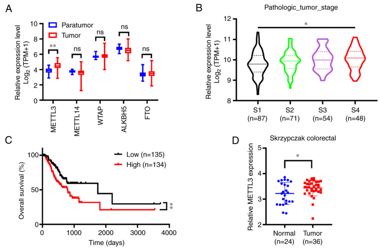
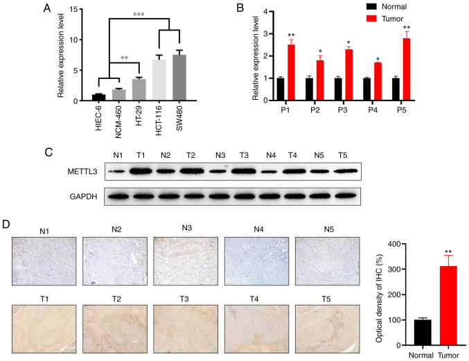
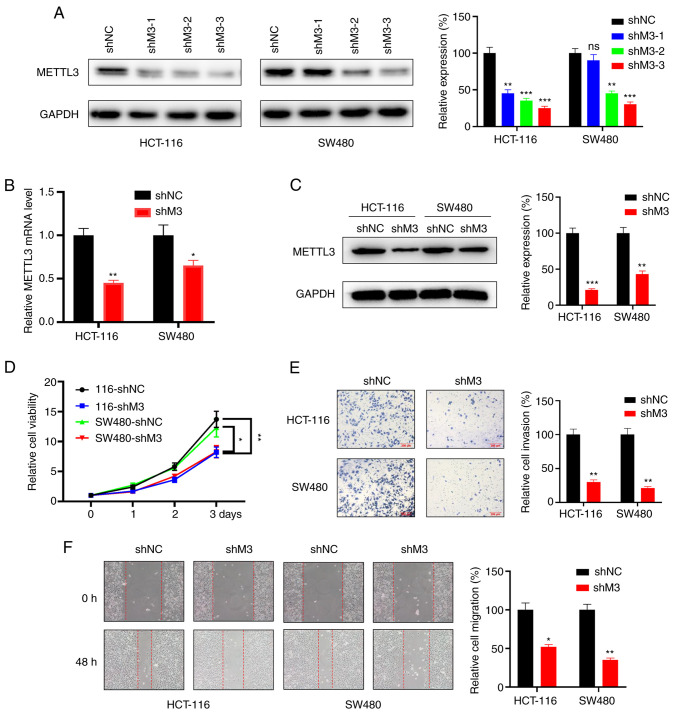
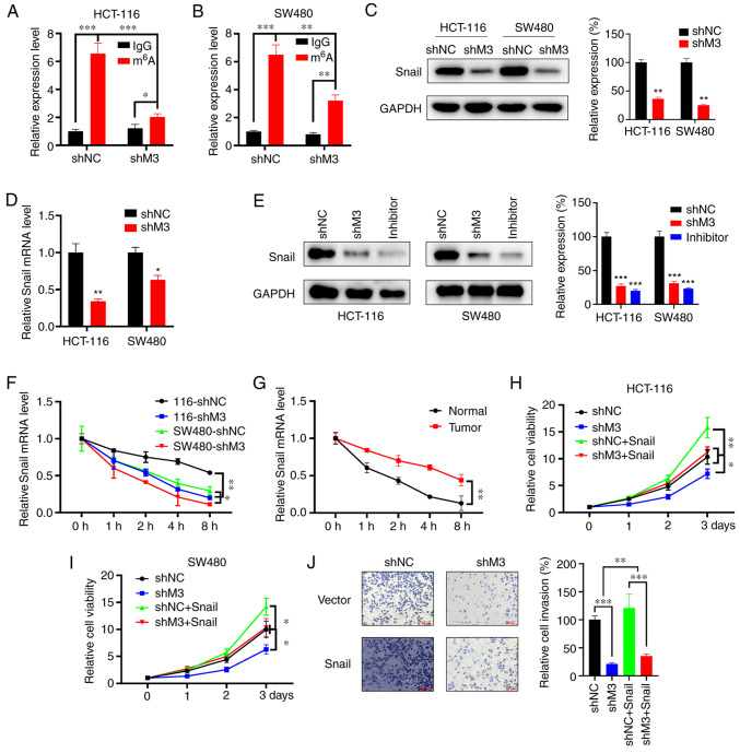
Nitrogen 6-methyladenosine (m6A) is the result of methylation of nitrogen-6 on adenosine, and is the most abundant chemical modification of eukaryotic mRNA. Dysregulation of m6A methylation has been implicated in cancer development and progression through various mechanisms. This type of methylation is primarily regulated by methyltransferase-like 3 (METTL3). However, the molecular mechanisms underlying the role of METTL3 in colorectal cancer (CRC) have not been extensively elucidated. The present study explored m6A modification and the underlying mechanism of m6A, which serve regulatory roles in the development of CRC. It was found that METTL3 is upregulated in CRC cell lines and tissues, and its expression positively correlated with poor overall survival (OS). Mechanistically, the present study demonstrated that METTL3 methylates Snail mRNA, thus stabilizing it to promote CRC malignancy. The present findings indicate that m6A modification is involved in CRC tumorigenesis, and highlight its potential as a therapeutic target against CRC.

Keywords: Snail; colorectal cancer; methyltransferase-like 3; nitrogen 6-methyladenosine.


Conflict of interest statement

The authors declare that they have no competing interests.

Figures

Figure 1.
Figure 1.
METTL3 is significantly upregulated in CRC on TCGA and Oncomine databases. (A) Nitrogen 6-methyladenosine-related enzymes mRNA expression levels in CRC vs. normal tissues according to TCGA database. (B) METTL3 expression levels in different clinical stages of CRC exhibited the following order: Clinical stages 1, 2, 3 and 4, as represented by the violin plots. (C) The overall survival of patients with CRC who exhibited high (n=135) vs. low (n=134) levels of METTL3 was plotted by the Kaplan-Meier method. (D) METTL3 mRNA expression levels in CRC and normal tissues according to the Oncomine database ‘Skrzypczak colorectal’. Error bars represent standard deviation. *P<0.05, **P<0.01. METTL3, methyltransferase-like 3; CRC, colorectal cancer; TCGA, The Cancer Genome Atlas; TPM, transcript count per million; ns, not significant; WTAP, Wilms' tumor 1-associating protein; ALKBH5, α-ketoglutarate-dependent dioxygenase AlkB homolog 5; FTO, fat mass and obesity-associated protein.
Figure 2.
Figure 2.
METTL3 is significantly upregulated in CRC tissues compared with its expression levels in the corresponding adjacent normal colon tissues. (A) METTL3 mRNA expression levels in the human CRC cell lines, HT-29, HCT-116 and SW480, the normal cell lines NCM-460 and HIEC-6. (B) Relative mRNA expression levels of METTL3 in CRC tissues compared with those in the corresponding adjacent normal tissues (n=5). (C) Protein expression of METTL3 in CRC vs. the corresponding adjacent normal tissues. GAPDH was used as a loading control. The right panel shows the quantitative analysis of the western blot results, as analyzed by paired Student's t-test. (D) The expression of METTL3 in CRC and adjacent normal tissues was detected by IHC staining (magnification, ×100; n=5). Error bars represent standard deviation. *P<0.05, **P<0.01, ***P<0.001. METTL3, methyltransferase-like 3; CRC, colorectal cancer; P, patient; N, normal; T, tumor.
Figure 3.
Figure 3.
M3 knockdown represses cell proliferation, invasion and migration in vitro. (A) Protein levels (right, quantitative analysis) of M3 in HCT-116 and SW480 cells upon transfection with a scrambled shRNA or with one of three different M3-silencing shRNAs. (B) mRNA levels of M3 in HCT-116 and SW480 cells infected with lentivirus carrying the indicated shRNA. (C) Protein levels of M3 in HCT-116 and SW480 cells infected with lentivirus carrying the indicated shRNA (right, quantitative analysis). (D) The proliferation rate of stable scrambled shRNA (shNC) and stable METTL3 knockdown (shM3) cells was evaluated by Cell Counting Kit-8 assay in HCT-116 and SW480 cells. (E) Transwell assay (right, quantitative analysis) was employed to analyze the difference in the invasion ability of shNC and shM3 cells. (F) Wound healing assay (magnification, ×200; right, quantitative analysis) revealed differences in the migration ability of shNC and shM3 cells. Each experiment was repeated three times independently. Error bars represent standard deviation. *P<0.05, **P<0.01, ***P<0.001. M3, methyltransferase-like 3; sh, small hairpin RNA; NC, negative control; ns, not significant.
Figure 4.
Figure 4.
Snail mediates M3-induced malignancy in CRC. m6A RIP-qPCR analysis of Snai1 mRNA in shNC- and shM3-infected (A) HCT-116 and (B) SW480 cells. (C) Snail protein expression levels (right, quantitative analysis) in stable with/without M3 knockdown HCT-116 and SW480 cells. (D) Snail mRNA expression levels in stably infected HCT-116 and SW480 cells with/without M3 knockdown. (E) Wild-type cells were treated with S-adenosylhomocysteine at a final concentration of 100 nM for 48 h, followed by detection of Snail protein expression (right, quantitative analysis) using western blot analysis. shNC and shM3 cells were included for analysis. (F) Snail mRNA expression levels in shNC and shM3 cells treated with Act-D (5 µg/ml) for the indicated time points were evaluated by qPCR. (G) Snail mRNA expression levels in CRC and adjacent normal tissue cells treated with Act-D (5 µg/ml) for the indicated time points were evaluated by qPCR. The proliferation rate of (H) HCT-116 and (I) SW480 cells transfected with shNC and shM3 and/or overexpressing Snail was evaluated using a Cell Counting Kit-8 assay at the indicated time points. (J) shNC or shM3 cells transiently overexpressing Snail or pcDNA3 vector were subjected to invasion assays for 24 h and subsequently analyzed with CytoSelect™ 24-well Cell Invasion assay kits (magnification, ×100; 8 µm; left). The invaded cells were then quantitatively analyzed (right). All experiments were performed in triplicate. Error bars represent standard deviation. *P<0.05, **P<0.01, ***P<0.001. M3, methyltransferase-like 3; CRC, colorectal cancer; m6A, nitrogen 6-methyladenosine; RIP, RNA immunoprecipitation; qPCR, quantitative PCR; sh, small hairpin RNA; NC, negative control; Act-D, actinomycin D.

Similar articles

Cited by

References

    1. Siegel RL, Miller KD, Fedewa SA, Ahnen DJ, Meester RG, Barzi A, Jemal A. Colorectal cancer statistics, 2017. CA Cancer J Clin. 2017;67:177–193. doi: 10.3322/caac.21395. - DOI - PubMed
    1. National Health Commission of The People's Republic of China, corp-author. National guidelines for diagnosis and treatment of colorectal cancer 2020 in China (English version) Chin J Cancer Res. 2020;32:415–445. doi: 10.21147/j.issn.1000-9604.2020.04.01. - DOI - PMC - PubMed
    1. Degiuli M, Reddavid R, Ricceri F, Di Candido F, Ortenzi M, Elmore U, Belluco C, Rosati R, Guerrieri M, Spinelli A, Members of the Italian Society of Surgical Oncology Colorectal Cancer Network (SICO-CCN) Collaborative Group Segmental colonic resection is a safe and effective treatment option for colon cancer of the splenic flexure: A nationwide retrospective study of the italian society of surgical oncology-colorectal cancer network collaborative group. Dis Colon Rectum. 2020;63:1372–1382. doi: 10.1097/DCR.0000000000001743. - DOI - PubMed
    1. Tang R, Cheng AJ, Wang JY, Wang TC. Close correlation between telomerase expression and adenomatous polyp progression in multistep colorectal carcinogenesis. Cancer Res. 1998;58:4052–4054. - PubMed
    1. Pichler M, Stiegelbauer V, Vychytilova-Faltejskova P, Ivan C, Ling H, Winter E, Zhang X, Goblirsch M, Wulf-Goldenberg A, Ohtsuka M, et al. Genome-Wide miRNA analysis identifies miR-188-3p as a novel prognostic marker and molecular factor involved in colorectal carcinogenesis. Clin Cancer Res. 2017;23:1323–1333. doi: 10.1158/1078-0432.CCR-16-0497. - DOI - PMC - PubMed