. 2021 Aug 17:9:e12000.
doi: 10.7717/peerj.12000. eCollection 2021.

Virulence effector SidJ evolution in Legionella pneumophila is driven by positive selection and intragenic recombination

Affiliations

Virulence effector SidJ evolution in Legionella pneumophila is driven by positive selection and intragenic recombination

Xiao-Yong Zhan et al. PeerJ. .

Abstract

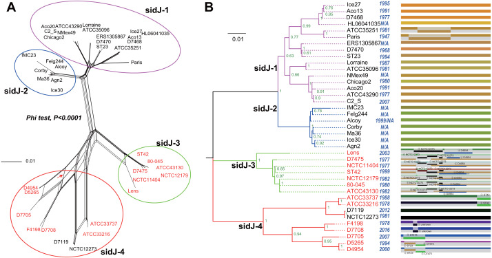
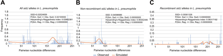
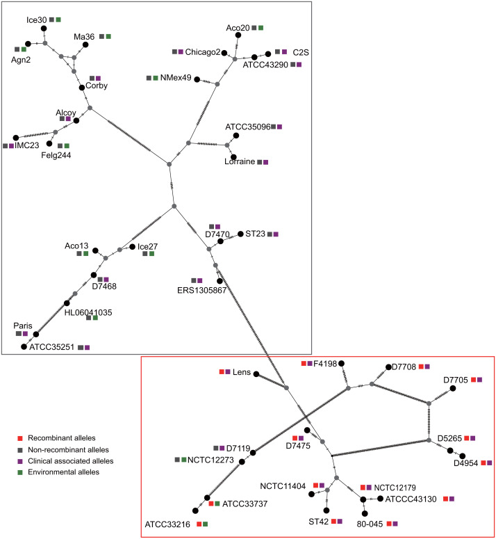
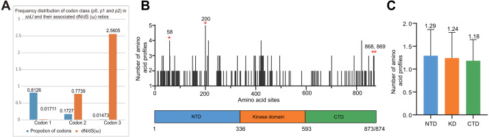
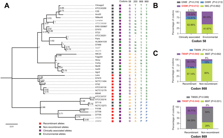
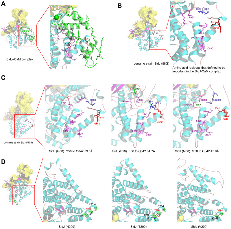
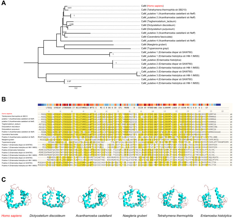
Effector proteins translocated by the Dot/Icm type IV secretion system determine the virulence of Legionella pneumophila (L. pneumophila). Among these effectors, members of the SidE family (SidEs) regulate several cellular processes through a unique phosphoribosyl ubiquitination mechanism mediated by another effector, SidJ. Host-cell calmodulin (CaM) activates SidJ to glutamylate the SidEs of ubiquitin (Ub) ligases and to make a balanced Ub ligase activity. Given the central role of SidJ in this regulatory process, studying the nature of evolution of sidJ is important to understand the virulence of L. pneumophila and the interaction between the bacteria and its hosts. By studying sidJ from a large number of L. pneumophila strains and using various molecular evolution algorithms, we demonstrated that intragenic recombination drove the evolution of sidJ and contributed to sidJ diversification. Additionally, we showed that four codons of sidJ which are located in the N-terminal (NTD) (codons 58 and 200) and C-terminal (CTD) (codons 868 and 869) domains, but not in the kinase domain (KD) had been subjected to strong positive selection pressure, and variable mutation profiles of these codons were identified. Protein structural modeling of SidJ provided possible explanations for these mutations. Codons 868 and 869 mutations might engage in regulating the interactions of SidJ with CaM through hydrogen bonds and affect the CaM docking to SidJ. Mutation in codon 58 of SidJ might affect the distribution of main-chain atoms that are associated with the interaction with CaM. In contrast, mutations in codon 200 might influence the α-helix stability in the NTD. These mutations might be important to balance Ub ligase activity for different L. pneumophila hosts. This study first reported that intragenic recombination and positive Darwinian selection both shaped the genetic plasticity of sidJ, contributing to a deeper understanding of the adaptive mechanisms of this intracellular bacterium to different hosts.

Keywords: Adaptive evolution; Evolution; Intragenic recombination; Legionella pneumophila; Positive selection; SidJ; Virulence effector.


Conflict of interest statement

The authors declare there are no competing interests.

Figures

Figure 1
Figure 1. Phylogenetic relationships among L. pneumophila sidJ alleles.
(A) Neighbor-net phylogenetic network shows the relationship among 39 sidJ alleles (see Table S1). The sidJ-1 subgroup is shown in a purple cycle, sidJ-2 blue, sidJ-3 green, and sidJ-4 red. The internal nodes represent hypothetical ancestral alleles and edges correspond to reticulate events such as recombination in the evolution of sidJ. The red arrow points to a representative reticulate event. (B) The evolutionary history of sidJ was inferred by using the Maximum Likelihood method. The ML tree was constructed from the alignment of nucleotide sequences of 39 alleles. Allele names were marked as their representative strain names. The earliest possible year for the allele to arose is shown in blue. Numbers on the interior branches represent bootstrap values and are indicated when the values are >0.5. The tree is drawn to scale, with branch lengths measuring in the number of substitutions per site. Branches in the same color were clustered into a group. Allele names are marked in red to indicate that they are recombinants. Unique recombination events detected by six recombination detection methods implemented under the RDP4 based on sidJ alleles are mapped onto the corresponding breaking point positions in the alignment (in the right of the figure). Recombination events that were identified by four or more methods were selected and numbered according to the RDP4 analysis, and the minor parent names of the recombinant alleles are shown nearby the breaking point positions (see Table 1).
Figure 2
Figure 2. Frequency distribution of the number of pairwise nucleotide differences (mismatch) between sidJ alleles (haplotypes).
(A) Mismatch distribution for the total data set (39 alleles); and the two allele groups including the (B) non-recombinant alleles and the (C) recombinant alleles are indicated. The solid orange line is the theoretical distribution under the assumption of population expansion.
Figure 3
Figure 3. Parsimony (TCS) network of L. pneumophila sidJ haplotypes (alleles).
The alleles were obtained from 116 worldwide isolates. Each oblique line between haplotypes (haplotype name is shown as its representative isolate name) represents one mutational difference. The connections are mutational steps between individuals. Unlabeled nodes (gray circles) indicate inferred steps that have not been found in the sampled populations as of yet. Boxes indicate major haplotype groups. Most haplotypes included in red dotted boxes are the recombinant ones, while those included in gray boxes are non-recombinant ones.
Figure 4
Figure 4. Frequency distribution of codon classes of sidJ, and domain architecture and amino acid profiles of each domain in the SidJ.
(A) Frequency distribution of codon classes (p0 = negative selection, p1 = neutral selection, and p2 = positive selection) and their associated dN/dS (ω) ratios under M3 model. (B) Domain architecture of SidJ depicting the NTD (blue), the KD (orange), and the CTD (green). Amino acid profiles of each site are indicated. Positive selection sites are marked in red, and the number of their amino acid profiles is shown. (C) An average number of amino acid profiles in each domain. Data are shown in mean and standard deviation.
Figure 5
Figure 5. Phylogenetic relationships among SidJ proteins of L. pneumophila from different sources, and with different mutation profiles at positive selection sites.
(A) Numbers on the interior branches represent bootstrap values and are indicated when the values are > 0.5. Protein names were marked as their representative allele (strain) names. Alleles with the same protein sequences were marked in gray. (B) Composition of different mutation profiles of positive selection sites in the clinically associated and environmental SidJ, or (C) in the recombinants and non-recombinants. Comparison of frequencies of codons between different groups was carried out by using the Chi-square tests or Fisher’s Exact tests.
Figure 6
Figure 6. Structure of SidJ and potential influence of mutation in positive selection sites.
The yellow covering shows the surface of the KD domain of SidJ. The secondary structure elements of SidJ are colored in cyan (helix), purple (sheet), and orange (loop) respectively. (A) Structure of the SidJ-CaM complex (PDB ID: 6K4K). The CaM is shown in green. The amino acid residues are shown as sticks. Purple sticks indicate a part of representative amino acid residues of SidJ that interact with CaM amino acid residues (gray sticks). Red sticks indicate the IQ motif (I841Q842) of SidJ. Hydrogen bonds (colored in red) including S808(SidJ) and E812(SidJ): R38(CaM) are shown. (B) The overall structure of the Lorraine strain SidJ. The relative position of amino acid residues that are defined to be important in interacting with CaM residues are shown as purple sticks and the IQ motif (I841Q842) is shown in red. (C) Mutation of positive selection site (codon 58) causes a distance change of closest atoms between the core of IQ motif which is crucial in SidJ-CaM binding. (D) The number of hydrogen bonds (colored in red) vary among different mutation profiles of codon 200.
Figure 7
Figure 7. CaM is variable among potential L. penumophila hosts.
(A) Phylogenetic relationship among CaM from Homo sapiens and other potentialL. penumophila environmental hosts. (B) Variability of CaM protein sequences among Homo sapiens and other potential L. penumophila environmental hosts. Amino acid residues marked in red indicate that 50% of the protein harbor the same residue in this site. (C) CaM protein structure comparisons among six potential L. penumophila hosts. The evolutionary history of CaM protein was inferred by using the Maximum Likelihood method and LG model. Bootstrap values were estimated using 1,000 replications. Numbers on the interior branches represent bootstrap values and are indicated when the values are > 0.5. Details of CaM from Homo sapiens and other potentialL. penumophila environmental hosts are shown in Table S6. We utilized the CaM protein with the NCBI accession number AAD45181.1 as the representative for Homo Sapiens, AAA33172.1 as the representative for Dictyostlium discoideum, XP_004334690.1 as the representative for Acanthamoeba castellanii, XP_002674748.1 as the representative for Naegleria gruberi, XP_001022775.2 as the representative for Tetrahymena thermophila, and XP_651708.1 as the representative for Entamoeba histolytica. All three-dimensional structures of CaM are shown in the same visual angle.

Similar articles

Cited by

References

    1. Al-Khodor S, Price CT, Habyarimana F, Kalia A, Abu Kwaik Y. A Dot/Icm-translocated ankyrin protein of Legionella pneumophila is required for intracellular proliferation within human macrophages and protozoa. Molecular Microbiology. 2008;70:908–923. doi: 10.1111/j.1365-2958.2008.06453.x. - DOI - PMC - PubMed
    1. Albert-Weissenberger C, Cazalet C, Buchrieser C. Legionella pneumophila —a human pathogen that co-evolved with fresh water protozoa. Cellular & Molecular Life Ences. 2007;64:432. doi: 10.1007/s00018-006-6391-1. - DOI - PMC - PubMed
    1. Anand IS, Choi W, Isberg RR. Components of the endocytic and recycling trafficking pathways interfere with the integrity of the Legionella-containing vacuole. Cellular Microbiology. 2020;22:e13151. doi: 10.1111/cmi.13151. - DOI - PMC - PubMed
    1. Anisimova M, Nielsen R, Yang Z. Effect of recombination on the accuracy of the likelihood method for detecting positive selection at amino acid sites. Genetics. 2003;164:1229–1236. doi: 10.1093/genetics/164.3.1229. - DOI - PMC - PubMed
    1. Bhogaraju S, Bonn F, Mukherjee R, Adams M, Pfleiderer MM, Galej WP, Matkovic V, Lopez-Mosqueda J, Kalayil S, Shin D, Dikic I. Inhibition of bacterial ubiquitin ligases by SidJ-calmodulin catalysed glutamylation. Nature. 2019;572:382–386. doi: 10.1038/s41586-019-1440-8. - DOI - PMC - PubMed

LinkOut - more resources