

Imaging update in spondyloarthropathy
- PMID: 34458093
- PMCID: PMC8379506
- DOI: 10.1016/j.jcot.2021.101564
Imaging update in spondyloarthropathy
Abstract
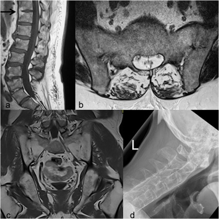
Although our understanding of axial spondyloarthropathy (axSpA) has increased recently, there has not been a concurrent improvement in patient diagnosis with delays contributing to patient morbidity. Imaging findings of axSpA can be subtle and may be dismissed often due to lack of understanding by reporters and importantly clinicians who do not suspect the disease. Recognition of the importance of imaging has led to the inclusion of MRI as part of the diagnostic criteria for axSpA. With this in mind, a number of advancements have been made in an attempt to increase our diagnostic accuracy on imaging. This article will give an overview of these techniques as well as a recap of the imaging features of axSpA.
Keywords: Inflammatory lesions; Rheumatology; Spondyloarthropathy; Structural lesions.
© 2021 Delhi Orthopedic Association. All rights reserved.
Conflict of interest statement
Nil.
Figures


Similar articles
-
The Fascinating Paradox of Osteoporosis in Axial Spondyloarthropathy.J Rheumatol. 2017 Dec;44(12):1767-1776. doi: 10.3899/jrheum.170051. Epub 2017 Oct 1. J Rheumatol. 2017. PMID: 28966207 Review.
-
Evaluation of the performances of 'typical' imaging abnormalities of axial spondyloarthritis: results of the cross-sectional ILOS-DESIR study.RMD Open. 2019 May 28;5(1):e000918. doi: 10.1136/rmdopen-2019-000918. eCollection 2019. RMD Open. 2019. PMID: 31245053 Free PMC article.
-
Diagnostic utility of whole spine and thoracic spine MRI corner inflammatory lesions in axial spondyloarthritis.Ther Adv Musculoskelet Dis. 2020 Nov 24;12:1759720X20973922. doi: 10.1177/1759720X20973922. eCollection 2020. Ther Adv Musculoskelet Dis. 2020. PMID: 33281954 Free PMC article.
-
Effects of ankylosing spondylitis and non-radiographic axial spondyloarthropathy on female sexual functions.Clin Exp Rheumatol. 2022 May;40(5):967-974. doi: 10.55563/clinexprheumatol/raqpga. Epub 2021 Jun 8. Clin Exp Rheumatol. 2022. PMID: 34128800
-
Therapeutic exercises and rehabilitation in axial spondyloarthropathy: Balancing benefits with unique challenges in the Asia-Pacific countries.Int J Rheum Dis. 2021 Feb;24(2):170-182. doi: 10.1111/1756-185X.14035. Epub 2020 Nov 27. Int J Rheum Dis. 2021. PMID: 33244895 Review.
Cited by
-
Updates of the imaging of musculoskeletal problems.J Clin Orthop Trauma. 2021 Sep 24;22:101612. doi: 10.1016/j.jcot.2021.101612. eCollection 2021 Nov. J Clin Orthop Trauma. 2021. PMID: 34631415 Free PMC article. No abstract available.
-
Vertebral Related Diseases in Healthcare: The Role of Pain Management and Rehabilitation.Healthcare (Basel). 2022 Jun 14;10(6):1109. doi: 10.3390/healthcare10061109. Healthcare (Basel). 2022. PMID: 35742160 Free PMC article.
References
-
- Pathak H., Gaffney K. Spectrum of spondyloarthritis. Indian J Rheumatol. 2020;15(Suppl S1):34–39.
-
- Shah A., Paramesparan K., Rennie W.J. Peripheral seronegative spondyloarthritis – updates on critical criteria. Indian J Musculoskelet Radiol. 2019;1(2):101–107.
-
- Jain, N., Byravan, S., Stairs, J., Rennie, W.J., Moorthy, A. Rheum, Vol 60(S-1).
-
- Ritchlin C., Adamopoulos I.E. Axial spondyloarthritis: new advances in diagnosis and management BMJ. 2021;372 - PubMed
-
- Chen M., Elawad A., Herregonds N., Jans L., Rennie W.J. Cutting edge technologies in the imaging of spondyloarthritis. Indian J Rheumatol. 2020;15(Suppl S1):27–33.
LinkOut - more resources
Full Text Sources