Review
. 2021 Aug 12:9:701950.
doi: 10.3389/fcell.2021.701950. eCollection 2021.

Epidermal Lamellar Body Biogenesis: Insight Into the Roles of Golgi and Lysosomes

Affiliations
Review

Epidermal Lamellar Body Biogenesis: Insight Into the Roles of Golgi and Lysosomes

Sarmistha Mahanty et al. Front Cell Dev Biol. .

Abstract

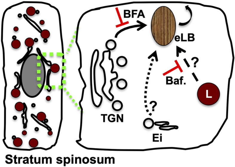
Epidermal lamellar bodies (eLBs) are secretory organelles that carry a wide variety of secretory cargo required for skin homeostasis. eLBs belong to the class of lysosome-related organelles (LROs), which are cell-type-specific organelles that perform diverse functions. The formation of eLBs is thought to be related to that of other LROs, which are formed either through the gradual maturation of Golgi/endosomal precursors or by the conversion of conventional lysosomes. Current evidence suggests that eLB biogenesis presumably initiate from trans-Golgi network and receive cargo from endosomes, and also acquire lysosome characteristics during maturation. These multistep biogenesis processes are frequently disrupted in human skin disorders. However, many gaps remain in our understanding of eLB biogenesis and their relationship to skin diseases. Here, we describe our current understanding on eLB biogenesis with a focus on cargo transport to this LRO and highlight key areas where future research is needed.

Keywords: Golgi; epidermal lamellar body; epidermis; lysosome; lysosome-related organelle.


Conflict of interest statement

The authors declare that the research was conducted in the absence of any commercial or financial relationships that could be construed as a potential conflict of interest.

Figures

FIGURE 1
FIGURE 1
Schematic representation of intracellular organelle dynamics in the epidermal sublayers. The human epidermis is composed of four functionally distinct sublayers formed by variable differentiation states of keratinocytes. Alterations in the extracellular composition and intracellular organelles of keratinocytes are represented according to the established studies (reviewed in Bikle et al., 2012). Stratum basale contains proliferative keratinocytes having all intracellular organelles similar to other cell types. These cells undergo differentiation (dotted arrow) after each cycle of asymmetric cell division and modify the Golgi organization and disperse lysosomes that result in the formation of the spinosum layer (Monteleon et al., 2018; Mahanty et al., 2019; Yamanishi et al., 2019). These cells are further subjected to late-stage differentiation and gradually lose their intracellular organelles while generating a new set of organelles called lamellar bodies (eLBs), which form the granulosum layer. These eLBs undergo exocytosis and generate the lipid sheets/barrier. Lastly, the cells of the granular layer undergo terminal differentiation, which forms SC (reviewed in Bikle et al., 2012). These cells are also called corneocytes (lack intracellular organelles) that remain embedded in lipid sheets produced by eLBs of the granular layer. The sequential changes in intracellular organelle dynamics, including the nucleus, are shown in the sublayers of skin (Akinduro et al., 2016). Extracellular ascending calcium gradient (Ca2+) and a descending nutrient gradient exist from the base of the epidermis. Note that the deformation of the nucleus initiates at the spinosum layer and eliminated through nucleophagy in the upper layers of the epidermis.
FIGURE 2
FIGURE 2
Modulation of Golgi or lysosome function alters the epidermal LB biogenesis and homeostasis. A set of TGN-derived vesicles possibly acts as the precursors of eLB in the differentiated keratinocytes of the spinosum layer, which matures into functional LBs post sequential maturation by receiving the cargo/membranes from the endolysosomal system (indicated as arrows). Several studies support this model: (a) inhibition of Golgi function with brefeldin A (BFA) blocks (shown as red color T) the production of epidermal lysosomes (L) (Mahanty et al., 2019) and LBs (Madison and Howard, 1996) and also inhibits keratinocyte differentiation (Mahanty et al., 2019); (b) inhibition of lysosome function by using bafilomycin (Baf.) causes defective epidermal architecture (shown as red color T) (Monteleon et al., 2018) and also inhibits the keratinocyte differentiation (Mahanty et al., 2019); and (c) silencing of Rab11A or knockout mouse of VPS33B or VIPAR displays defective eLB biogenesis, and barrier function suggests a role for Ei (consists of early/recycling endosomes) during their biogenesis (Reynier et al., 2016; Rogerson and Gissen, 2018). The drawn hypothetical model is based on the stepwise cargo transport to maturing TGN-derived precursor organelles, which generate functional eLBs. Differentiated keratinocyte of the stratum spinosum layer was shown on the left. The possible cargo trafficking routes to eLBs are indicated in the enhanced portion on the right side.
FIGURE 3
FIGURE 3
Proposed models for lipid matrix formation at the SC. Landmann’s model: In the upper spinosum and granular layer keratinocytes, eLBs are bud off from the TGN as discrete vesicles/tubules. The cargo in these vesicles is packaged in the form of disks that avoid the enzyme activity during their transport. These vesicles fuse with the plasma membrane of the granular cells and release the disks between the SG-SC junction, which merge together and generate the lipid sheets of the corneum layer (Landmann, 1986; Norlen, 2001a). Norlen’s model: eLBs are the continuous tubulo-reticular network from TGN to the plasma membrane and multilamellar lipid matrix at the SG-SC junction. Further, these organelles secrete lipids through the intersection-free unfoldings at the plasma membrane invaginations of the granular cells (Norlen, 2001a). In both these models, keratinocytes of the SG secrete eLBs (either in the form of vesicles or tubules), which form the SC layer.
FIGURE 4
FIGURE 4
Current models for the biogenesis of epidermal LBs. Based on the existing cargo/membrane transport routes to eLBs, we propose three variant mechanisms for the biogenesis of eLBs in the upper spinosum and granulosum layer keratinocytes. Model-1: LB precursors are derived from the TGN, which mature into functional eLBs upon receiving the cargo/membranes from early/recycling endosomes (Ei) and epidermal lysosomes (L) in a sequential manner. Model-2: TGN-derived LB precursors fuse with epidermal lysosomes and form hybrid organelles, which then mature into eLBs. The cargo transport from the early/recycling endosomes (Ei) to this hybrid organelles is not clear in this model. Model-3: The epidermal lysosomes of the spinosum layer mature into eLBs upon constant fusion of TGN-derived vesicles. As similar to model-2, the input from early/recycling endosomes (Ei) is not clear in this pathway. In all the three models, the matured eLBs fuse with the plasma membrane to deliver its content for the formation of lipid sheets at the SC.

References

    1. Akinduro O., Sully K., Patel A., Robinson D. J., Chikh A., Mcphail G., et al. (2016). Constitutive autophagy and nucleophagy during epidermal differentiation. J. Invest. Dermatol. 136 1460–1470. 10.1016/j.jid.2016.03.016 - DOI - PubMed
    1. Akiyama M. (2011). The roles of ABCA12 in keratinocyte differentiation and lipid barrier formation in the epidermis. Dermatoendocrinol 3 107–112. 10.4161/derm.3.2.15136 - DOI - PMC - PubMed
    1. Akiyama M. (2014). The roles of ABCA12 in epidermal lipid barrier formation and keratinocyte differentiation. Biochim. Biophys. Acta 1841 435–440. 10.1016/j.bbalip.2013.08.009 - DOI - PubMed
    1. Appelqvist H., Waster P., Eriksson I., Rosdahl I., Ollinger K. (2013). Lysosomal exocytosis and caspase-8-mediated apoptosis in UVA-irradiated keratinocytes. J. Cell Sci. 126 5578–5584. - PubMed
    1. Aymard E., Barruche V., Naves T., Bordes S., Closs B., Verdier M., et al. (2011). Autophagy in human keratinocytes: an early step of the differentiation? Exp. Dermatol. 20 263–268. 10.1111/j.1600-0625.2010.01157.x - DOI - PubMed

LinkOut - more resources