

Effects of Acute Diquat Poisoning on Liver Mitochondrial Apoptosis and Autophagy in Ducks
- PMID: 34458360
- PMCID: PMC8385319
- DOI: 10.3389/fvets.2021.727766
Effects of Acute Diquat Poisoning on Liver Mitochondrial Apoptosis and Autophagy in Ducks
Abstract
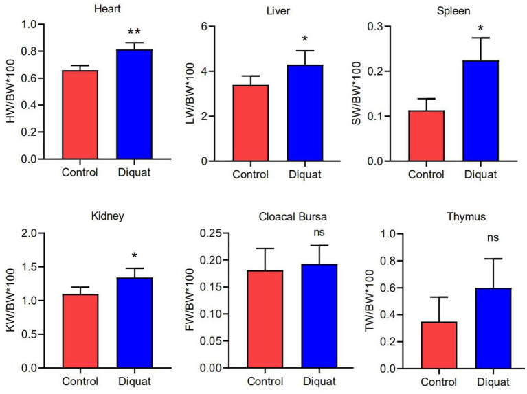
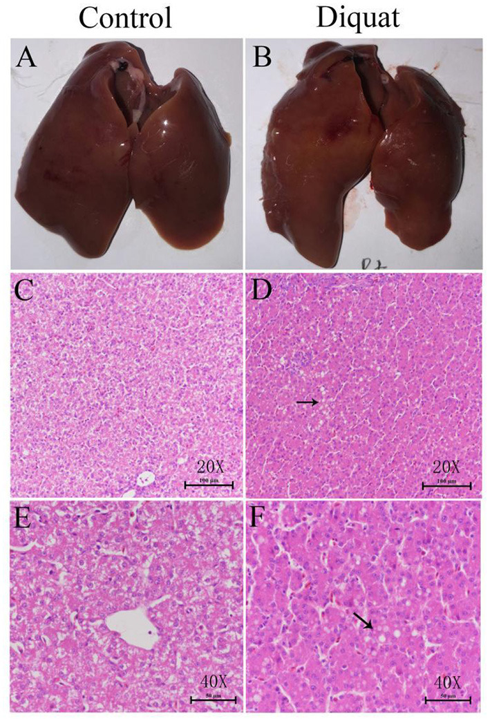
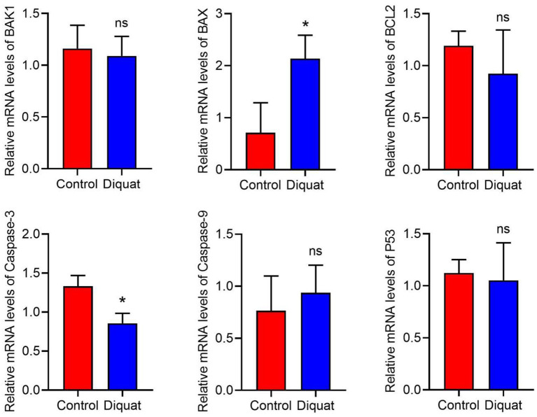
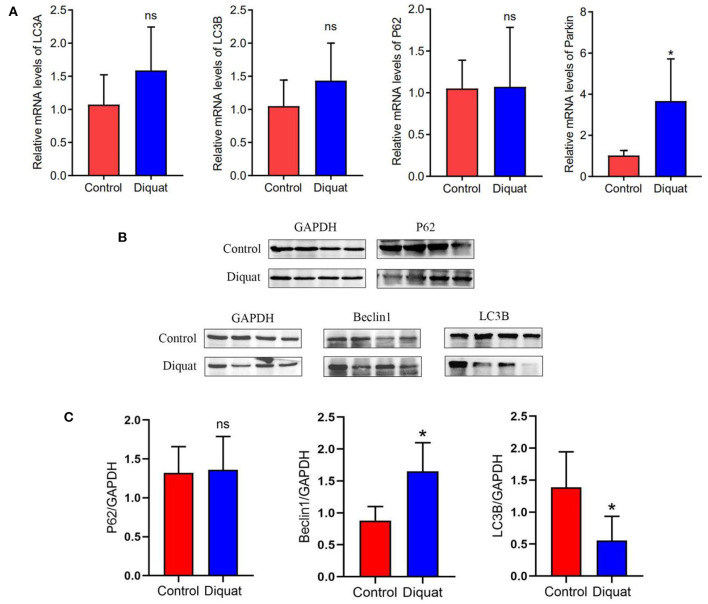
Diquat (DQ) is an effective herbicide and is widely used in agriculture. Due to persistent and frequent applications, it can enter into aquatic ecosystem and induce toxic effects to exposed aquatic animals. The residues of DQ via food chain accumulate in different tissues of exposed animals including humans and cause adverse toxic effects. Therefore, it is crucial and important to understand the mechanisms of toxic effects of DQ in exposed animals. We used ducks as test specimens to know the effects of acute DQ poisoning on mechanisms of apoptosis and autophagy in liver tissues. Results on comparison of various indexes of visceral organs including histopathological changes, apoptosis, autophagy-related genes, and protein expression indicated the adverse effects of DQ on the liver. The results of our experimental trial showed that DQ induces non-significant toxic effects on pro-apoptotic factors like BAX, BAK1, TNF-α, caspase series, and p53. The results revealed that anti-apoptotic gene Parkin was significantly upregulated, while an upward trend was also observed for Bcl2, suggesting that involvement of the anti-apoptotic factors in ducklings plays an important role in DQ poisoning. Results showed that DQ significantly increased the protein expression level of the autophagy factor Beclin 1 in the liver. Results on key autophagy factors like LC3A, LC3B, and p62 showed an upward trend at gene level, while the protein expression level of both LC3B and p62 reduced that might be associated with process of translation affected by the pro-apoptotic components such as apoptotic protease that inhibits the occurrence of autophagy while initiating cell apoptosis. The above results indicate that DQ can induce cell autophagy and apoptosis and the exposed organism may resist the toxic effects of DQ by increasing anti-apoptotic factors.
Keywords: apoptosis; autophagy; diquat; ducks; liver.
Copyright © 2021 Chen, Su, Lin, Lin, Shang, Hussain and Shi.
Conflict of interest statement
The authors declare that the research was conducted in the absence of any commercial or financial relationships that could be construed as a potential conflict of interest.
Figures
Similar articles
-
NF-κB/mTOR-mediated autophagy can regulate diquat-induced apoptosis.Arch Toxicol. 2019 May;93(5):1239-1253. doi: 10.1007/s00204-019-02424-7. Epub 2019 Mar 8. Arch Toxicol. 2019. PMID: 30848314
-
Pasteurella multocida causes liver injury in ducks by mediating inflammatory, apoptotic and autophagic pathways.Microb Pathog. 2023 Nov;184:106336. doi: 10.1016/j.micpath.2023.106336. Epub 2023 Sep 7. Microb Pathog. 2023. PMID: 37683832 Review.
-
Autophagy mediated FTH1 degradation activates gasdermin E dependent pyroptosis contributing to diquat induced kidney injury.Food Chem Toxicol. 2024 Feb;184:114411. doi: 10.1016/j.fct.2023.114411. Epub 2023 Dec 19. Food Chem Toxicol. 2024. PMID: 38128689
-
Autophagy inhibition uncovers the neurotoxic action of the antipsychotic drug olanzapine.Autophagy. 2014;10(12):2362-78. doi: 10.4161/15548627.2014.984270. Autophagy. 2014. PMID: 25551567 Free PMC article.
-
Imaging Findings and Toxicological Mechanisms of Nervous System Injury Caused by Diquat.Mol Neurobiol. 2024 Nov;61(11):9272-9283. doi: 10.1007/s12035-024-04172-x. Epub 2024 Apr 15. Mol Neurobiol. 2024. PMID: 38619744 Free PMC article. Review.
Cited by
-
Lethal diquat poisoning manifests as acute central nervous system injury and circulatory failure: A retrospective cohort study of 50 cases.EClinicalMedicine. 2022 Aug 11;52:101609. doi: 10.1016/j.eclinm.2022.101609. eCollection 2022 Oct. EClinicalMedicine. 2022. PMID: 35990582 Free PMC article.
-
Clinical and pathological characteristics of diquat poisoning-related acute kidney injury.Ren Fail. 2023;45(2):2283590. doi: 10.1080/0886022X.2023.2283590. Epub 2023 Nov 27. Ren Fail. 2023. PMID: 38010163 Free PMC article.
-
Effect of Diquat on gut health: molecular mechanisms, toxic effects, and protective strategies.Front Pharmacol. 2025 May 12;16:1562182. doi: 10.3389/fphar.2025.1562182. eCollection 2025. Front Pharmacol. 2025. PMID: 40421207 Free PMC article. Review.
-
Impact of Diquat on the Intestinal Health and the Composition and Function of the Gut Microbiome.Antioxidants (Basel). 2025 Jun 12;14(6):721. doi: 10.3390/antiox14060721. Antioxidants (Basel). 2025. PMID: 40563352 Free PMC article. Review.
-
Treatments with Diquat Reveal the Relationship between Protein Phosphatases (PP2A) and Oxidative Stress during Mitosis in Arabidopsis thaliana Root Meristems.Plants (Basel). 2024 Jul 10;13(14):1896. doi: 10.3390/plants13141896. Plants (Basel). 2024. PMID: 39065423 Free PMC article.
References
-
- Magalhães N, Carvalho F, Dinis-Oliveira RJ. Human and Experimental Toxicology of Diquat Poisoning: Toxicokinetics, Mechanisms of Toxicity, Clinical Features, and Treatment. London: SAGE Publications; (2018). - PubMed
LinkOut - more resources
Full Text Sources
Research Materials
Miscellaneous