. 2021 Aug 5;11(15):e4107.
doi: 10.21769/BioProtoc.4107.

Mouse Periosteal Cell Culture, in vitro Differentiation, and in vivo Transplantationin Tibial Fractures

Affiliations

Mouse Periosteal Cell Culture, in vitro Differentiation, and in vivo Transplantationin Tibial Fractures

Simon Perrin et al. Bio Protoc. .

Abstract

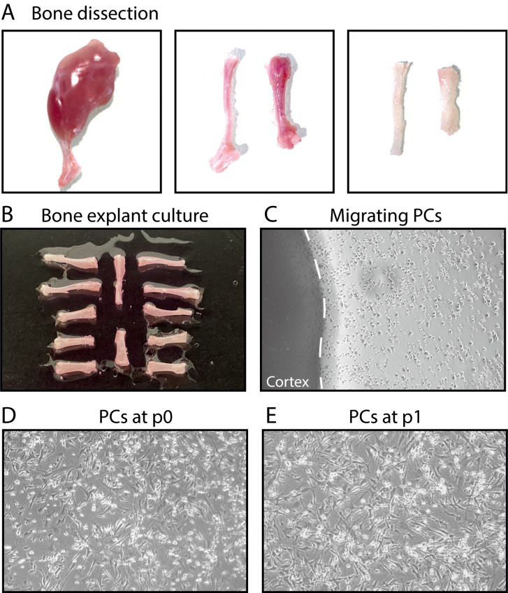
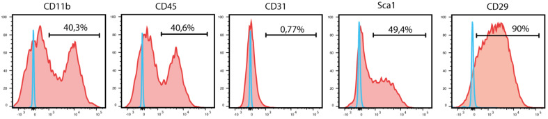
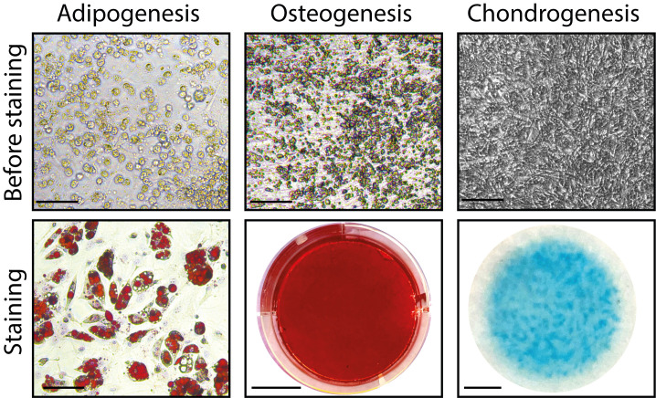
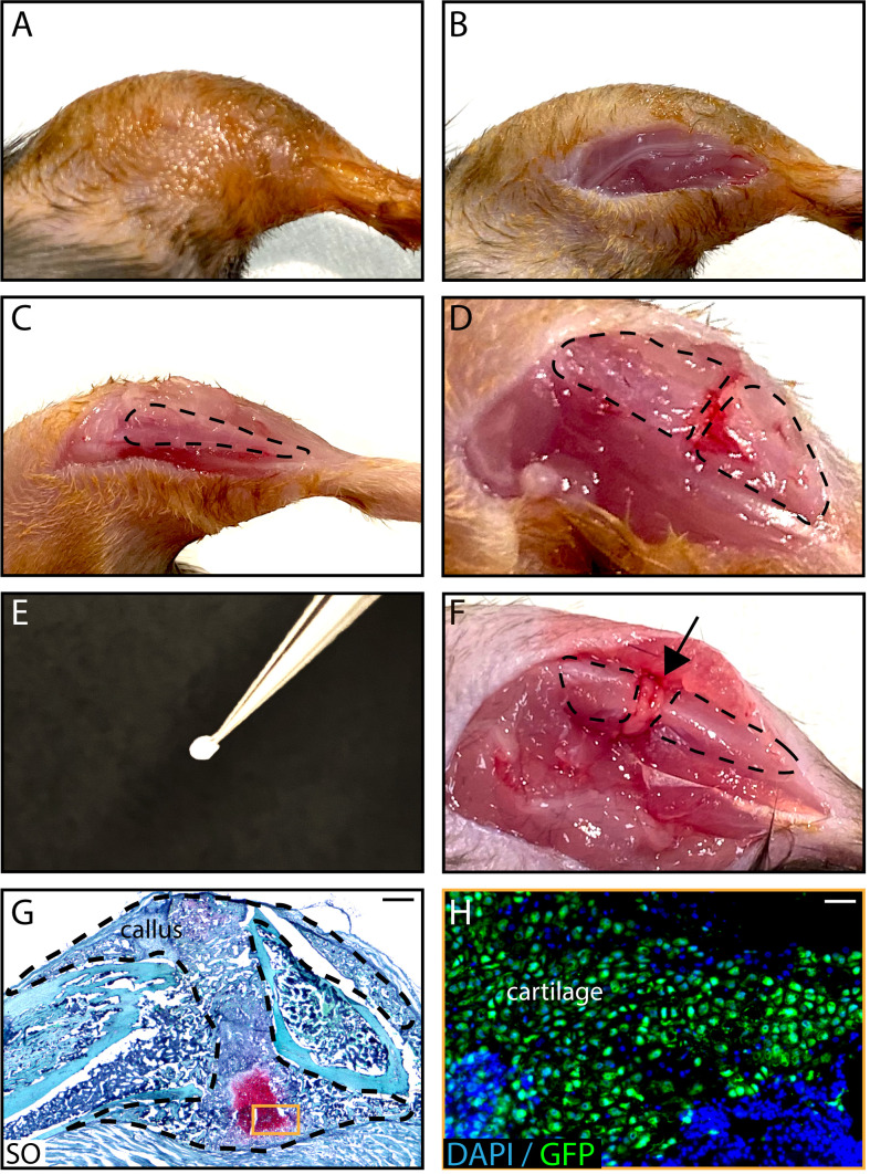
The periosteum covering the outer surface of bone contains skeletal stem/progenitor cells that can efficiently form cartilage and bone during bone repair. Several methods have been described to isolate periosteal cells based on bone scraping and/or enzymatic digestion. Here, we describe an explant culture method to isolate periosteum-derived stem/progenitor cells for subsequent in vitro and in vivo analyses. Periosteal cells (PCs) isolated using this protocol express mesenchymal markers, can be expanded in vitro, and exhibit high regenerative potential after in vivo transplantation at a fracture site, suggesting that this protocol can be employed for PC production to use in new cell-based therapies.

Keywords: Bone regeneration; In vitro differentiation; In vivo cell transplantation; Periosteum; Skeletal stem/progenitor cell.


Conflict of interest statement

Competing interestsThe authors declare no competing interests.

Figures

Figure 1.
Figure 1.. Steps of bone explant culture.
(A) Steps of bone dissection. Left, hindlimb free of skin. Middle, tibia, and femur free of muscle. Right, tibia and femur after cutting the epiphyses and flushing the bone marrow. (B) Tibias and femurs plated in a culture dish. (C) After a few days, periosteal cells (PCs) migrate out of the bone explants into the dish. (D-E) PCs at P0 and P1.
Figure 2.
Figure 2.. Flow cytometry analysis of periosteum-derived cells at P1.
Blue curves represent FMO controls, and red curves represent experimental samples.
Figure 3.
Figure 3.. In vitro adipogenic, osteogenic, and chondrogenic differentiation of PCs before and after staining.
Scale bars: before staining: 200 μm; staining: 50 μm (adipogenesis), 1 cm (osteogenesis), 2.5 mm (chondrogenesis).
Figure 4.
Figure 4.. In vivo transplantation of PCs isolated from GFP-actin donors at the fracture site of wild-type hosts
. After shaving and sanitizing the limb (A), an incision in the skin is performed (B) and the tibia is exposed (C, tibia is delineated by a dotted line) to induce a fracture (D). A Tisseel matrix pellet containing PCs (E) is transplanted at the fracture site (F, black arrow). (G) Representative image of a longitudinal callus section on day 14 post-fracture stained with SO. (H) GFP+ chondrocytes derived from PCs on an adjacent section of the callus. Scale bars: 1 mm (G) and 100 μm (H).

References

    1. Arnsdorf E. J., Luis M. J., Dennis R. C. and Christopher R. J.(2009). The periosteum as a cellular source for functional tissue engineering. Tissue Eng Part A 15(9): 2637-2642. - PMC - PubMed
    1. Arthur A. and Gronthos S.(2020). Clinical Application of Bone Marrow Mesenchymal Stem/Stromal Cells to Repair Skeletal Tissue. Int J Mol Sci 21(24): 9759. - PMC - PubMed
    1. Brownlow H. C., Reed A., Joyner C., Simpson A.H.(2000). Anatomical effects of periosteal elevation. J Orthop Res 18(3): 500-502. - PubMed
    1. Chang H. and Knothe Tate M. L.(2012) Concise review: the periosteum: tapping into a reservoir of clinically useful progenitor cells. Stem Cells Transl Med 1(6): 480-491. - PMC - PubMed
    1. Debnath S., Yallowitz A.R., McCormick J., Lalani S., Zhang T., Xu R., Li N., Liu Y. F., Yang Y. S., Eiseman M., Shim J. H., Hameed M., Healey J. H., Bostrom M. P., Landau D. A., Greenblatt M. B.(2018). Discovery of a periosteal stem cell mediating intramembranous bone formation. Nature 562(7725): 133-139. - PMC - PubMed

LinkOut - more resources